
A pediatric endocrinology clinic refers a 9-year-old with short stature for bone age, and orthopedics adds bilateral femorotibial length measurement for suspected leg-length difference. The child is anxious and parents ask about radiation and positioning. Which protocol correctly acquires bone age and long bone measurement images with calibration and minimal repeats?
Select an answer to reveal the explanation.
Short Explanation
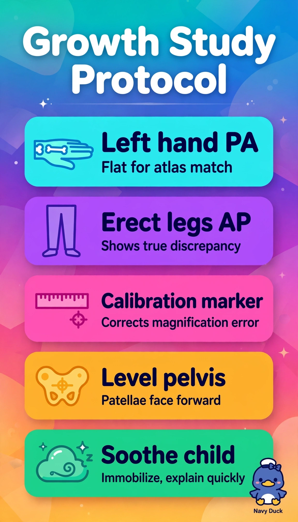
You're comparing growth to a standard atlas, not just taking a hand picture. A flat left hand and a calibrated standing leg view give true lengths, so left-hand PA plus measured erect legs is the accurate pair.
Full Explanation
Bone age compares ossification centers in the PA left hand and wrist to a standardized atlas, requiring a flat nonrotated PA projection of the non-dominant left hand to avoid magnification and rotation distortion, while long bone measurement requires erect AP views of the femorotibial segments with the pelvis level, patellae forward, and an external calibration marker at the level of the bone to correct for magnification and allow accurate length calculation. That combination standardizes both growth assessment and length difference with minimal exposure. An obliqued right hand distorts phalangeal centers and lacks atlas comparability, and supine femora without a marker and with outward rotation foreshorten length and hide discrepancy. A lateral hand superimposes carpals and cannot be graded, and recumbent lateral tibiae with a misplaced marker misrepresent weight-bearing length. Bilateral AP hands double dose without added value, and seated PA knees with the marker off anatomy eliminate calibration and functional length. Exam caveat: bone age is almost always left-hand PA unless injury or prior surgery dictates otherwise. Practical check: immobilize gently, include all digits and wrist, level the pelvis, and place the ruler at bone plane.