
A 250-bed hospital wants bedside abdominal images in a crowded ICU where transporting a Bucky is impractical and repeat rates are high from scatter fog. Engineering offers wireless flat-panel detectors and software scatter correction, with a choice of direct or indirect capture. Which detector and scatter strategy best balances bedside dose and contrast without a physical grid?
Select an answer to reveal the explanation.
Short Explanation
You solved portables elegantly. An indirect cesium panel plus smart grid software cleans scatter without wrestling a heavy grid at bedside.
Full Explanation
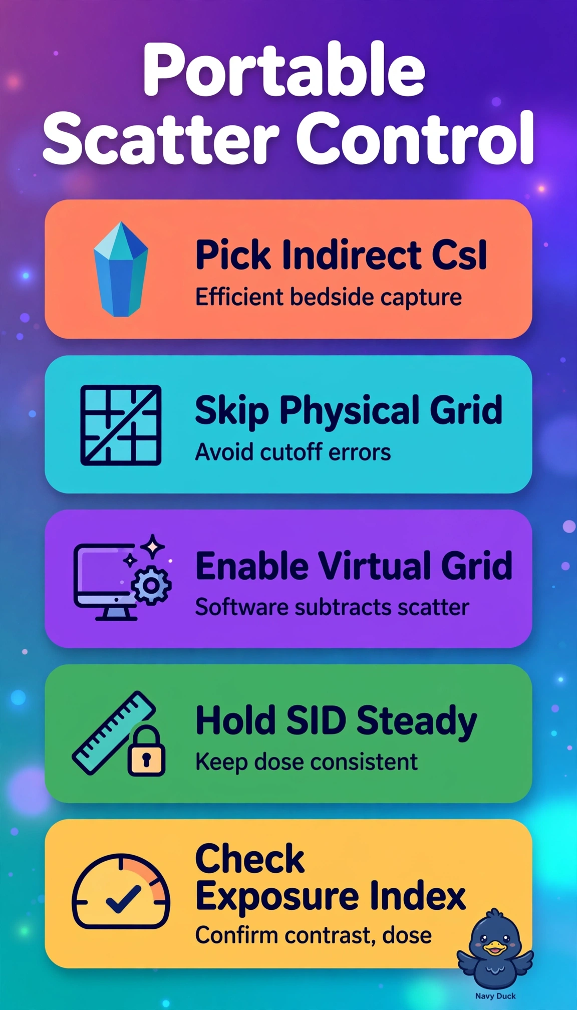
The correct pairing works because thick columnar CsI absorbs high-kVp abdominal photons efficiently and channels light to photodiodes with minimal blur, sustaining DQE and contrast at lower bedside dose, while virtual grid software models and subtracts scatter to cut fog-related repeats without grid weight, alignment, cutoff, or dose penalty in a crowded ICU. A direct a-Se detector with virtual software fails on capture physics because thin amorphous selenium captures poorly at abdominal energies, ideal for low-energy mammography resolution but noisy and dose-heavy for thick anatomy. A CsI panel using air-gap cleanup alone fails on bedside practicality because a diagnostic gap needs increased SID and space, causing magnification, motion blur and higher technique that crowded portable work cannot afford. A granular GOS indirect panel with virtual software fails on conversion efficiency because powder phosphor scatters light and yields lower MTF and DQE than needle CsI, demanding higher dose despite lower cost and ruggedness. Caveat for exams is that software improves contrast but never replaces tight collimation, correct kVp, and shielding, and higher DQE does not mean higher limiting resolution. Practical check is to verify wireless connection, virtual grid enabled, exposure index on target, and review scatter and positioning before repeating.