
A 68-year-old dysphagic stroke patient on a medical floor needs enteral feeding after an indwelling small-bore feeding tube was advanced at bedside. Aspiration pH is equivocal, and the physician orders a portable abdomen to confirm placement before feeding. The nurse asks if feeding can start. Which radiographic verification best confirms safe enteric placement before feeding?
Select an answer to reveal the explanation.
Short Explanation
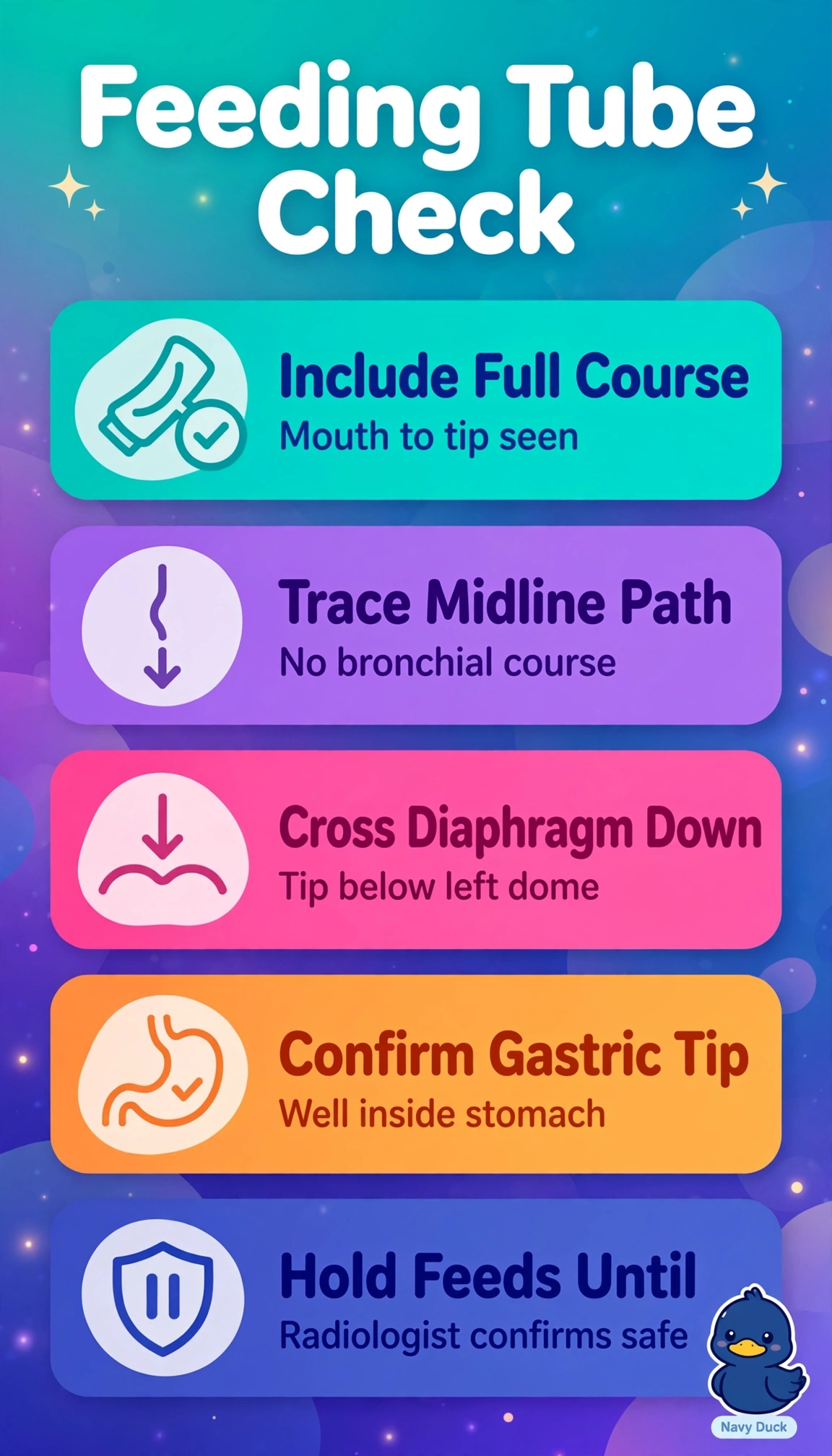
You are the last safety net before feeding. Seeing the tip clearly below the diaphragm in the stomach with a straight esophageal course proves safe placement.
Full Explanation
Safe feeding after blind small-bore placement in a dysphagic stroke patient with equivocal pH requires radiographic proof that the tip lies unequivocally in the gastric lumen well below the diaphragm and that the visible course remains midline through the esophagus without entering bronchus or lung, because small-caliber tubes can pass silently into the airway and dysphagia magnifies aspiration injury, so only that combined location-plus-course verification permits feeding. Confirming a tip just below the diaphragm at the gastroesophageal junction fails by location because ports at or above the lower esophageal sphincter deliver feed to distal esophagus or cardia with high reflux, vomiting and aspiration risk and does not meet the well-below criterion for gastric placement. Confirming a presumed gastric tip on a collimated abdominal view without tracing the thoracic segment fails by exclusion because without seeing midline descent through the thorax and midline diaphragmatic crossing one cannot rule out bronchial entry, looping, coiling or kinked passage that secondarily reaches the abdomen. Relying on an auscultated epigastric whoosh despite an apparently infra-diaphragmatic tip fails by method because insufflated air transmits widely and cannot distinguish gastric from esophageal, pleural or bronchial position, so guidelines require radiography when pH is equivocal or unobtainable. An exam caveat is that a portable abdomen cropping the lower chest is inadequate for initial confirmation and the image must include carina to upper abdomen with crisp tip visualization. A practical check is to trace the radiopaque line from pharynx downward, confirm midline esophageal passage, diaphragmatic crossing and leftward deviation into fundus or body, hold feeds, keep head elevated, and obtain formal radiology confirmation before flushing or feeding.