
A 78-year-old kyphotic patient with a suspected left hip fracture arrives on a stretcher, cannot stand or abduct the painful leg, has severe osteoporosis, and needs AP pelvis plus lateral hip views without worsening injury. Which trauma-adapted positioning modification should the radiographer apply to obtain diagnostic views safely?
Select an answer to reveal the explanation.
Short Explanation
You're working around pain, not through it, so bring the beam to the hip. Cross-table lateral is your side-step that keeps the hurt leg perfectly still.
Full Explanation
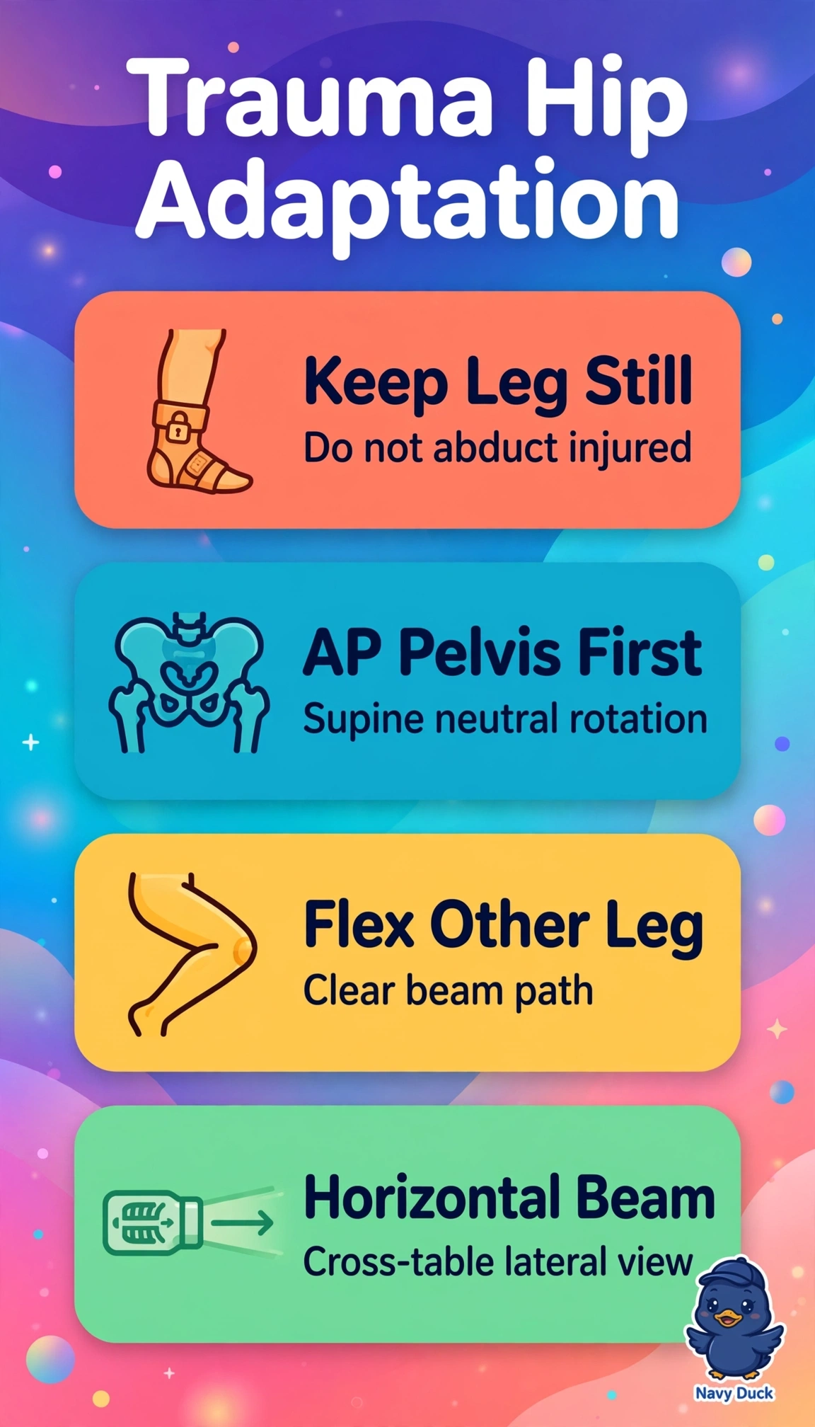
The trauma-adapted standard for this stretcher-bound, osteoporotic, kyphotic patient is supine AP pelvis with legs in neutral without manipulating the injured hip combined with horizontal-beam cross-table lateral, with the uninjured leg flexed and moved clear of the beam so the femoral head, neck and trochanters are visualized in two orthogonal planes without standing, abduction, transfer, or fracture displacement, while padding supports the kyphosis. Routine AP pelvis technique using bilateral 15-degree internal rotation fails by fracture-stability concept because rotating the suspected fractured femur to close the obturator foramina can displace an impacted femoral neck or intertrochanteric fracture and increase pain and bleeding. Forcing the spine flat for alignment fails by geriatric-positioning concept because fixed kyphosis requires stretcher-based imaging with head and knee supports, and flattening causes pain, respiratory compromise and motion without improving hip evaluation. Using only AP pelvis and AP hip without a lateral to reduce movement fails by orthogonal-view concept because displacement, angulation and femoral head-neck relationship cannot be assessed on frontal projection alone and surgical planning requires a true lateral. Exam caveat is to separate routine arthritis pelvis protocol with internal rotation and frog-leg lateral from suspected-fracture trauma protocol where neutral AP plus cross-table lateral is required. Practical check is to confirm the injured leg was never rotated or abducted, the uninjured leg is flexed clear, the cross-table beam is perpendicular to the femoral neck, and the lateral shows head-neck continuity before any transfer.