
An outpatient center repeats chest images because thin patients are overexposed and a scoliosis patient with hardware shows underexposure. The system uses three ionization chambers, center and outer detectors, and a backup timer. Which AEC setup and safety check best addresses detector selection and exposure termination?
Select an answer to reveal the explanation.
Short Explanation
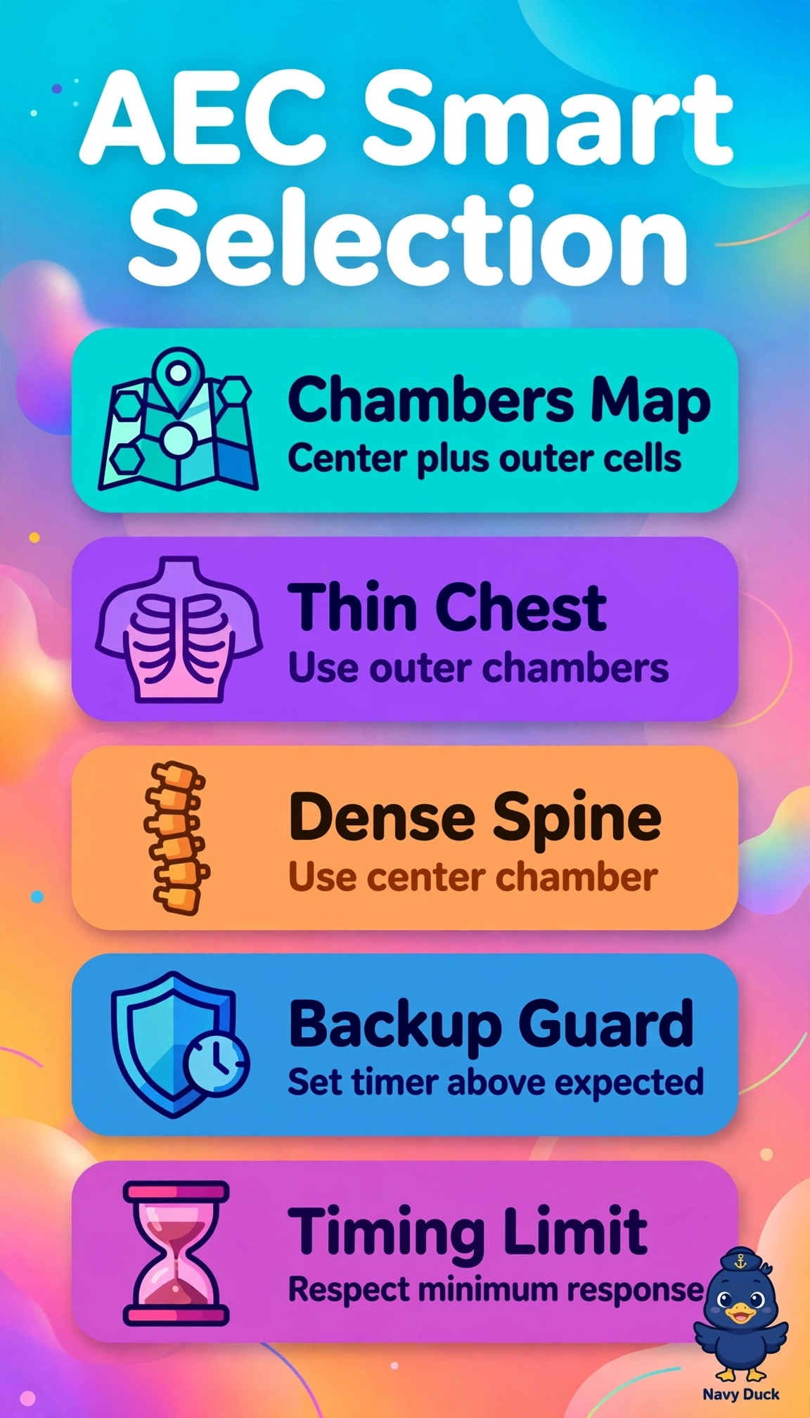
You’re matching the sensor to the anatomy like a spotlight. Choosing chambers under real lung or bone keeps density even because ionization chambers stop exposure on time.
Full Explanation
Correct setup matches detector position to tissue attenuation while preserving redundant termination because outer chambers lie under aerated lung in thin chests and terminate before mediastinal overpenetration causes overexposure, while the center chamber lies under the dense vertebral mass for scoliosis work and provides representative ionization when fully covered without hardware directly over it, and properly set backup mAs stops exposure if AEC cannot reach cutoff. A variant with correct chambers but backup set very high preserves geometry yet violates backup safety concept by permitting excessive dose and tube loading before termination, which defeats the purpose of redundant cutoff. A uniform center-chamber technique preserves backup safety and suits average chests yet violates anatomy-matching and minimum response concepts because the center detector behind thin mediastinum sees low attenuation and terminates late, reproducing thin-patient overexposure. Using outer chambers for the instrumented spine preserves thin-chest logic and backup yet violates representative sampling because off-center detectors under ribs, curvature, or metal edges do not sample the densest anatomy of interest, producing cutoff errors and the reported underexposure. Exam caveat is that avoiding hardware does not mean automatically selecting outer detectors; the selected detector must correspond to pathology and be fully covered by soft tissue without implant overlap. Practical check is to verify active detector lights, center anatomy over them, confirm kVp adequate for ionization chamber response, and set backup mAs at about one and one-half times expected manual mAs per technique chart.