
A 34-year-old emergency patient needs a supine AP abdomen to include pubic symphysis, kidneys, and lateral soft tissues, with correct mid-sagittal plane alignment and no rotation for obstruction workup. Which topographic landmark and central ray path should the radiographer use to include the required anatomy?
Select an answer to reveal the explanation.
Short Explanation
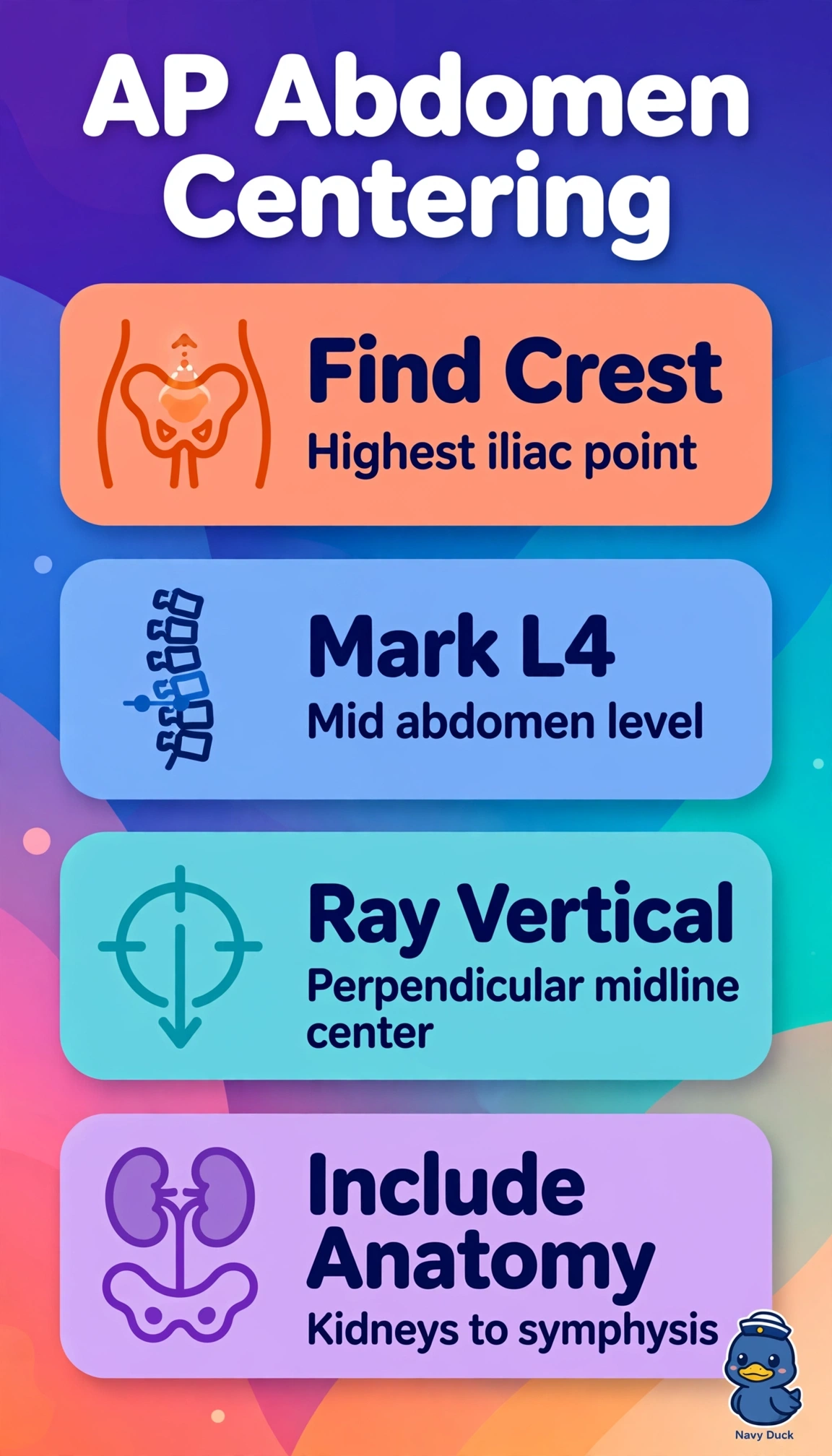
You’re framing the abdomen from kidneys to bladder in one shot, so aim for the middle. The iliac crest is your L4 landmark for that coverage.
Full Explanation
For a supine AP abdomen in an obstruction workup, include the superior renal poles through the pubic symphysis and the lateral flank soft tissues, with the midsagittal plane perpendicular to the detector and no rotation for symmetric bowel assessment. Centering midline at iliac crest level, around L4, with a vertical central ray places the beam midway between the superior and inferior limits; divergence helps capture the kidneys and symphysis together with even density and minimal distortion while preserving alignment. Centering at costal margin level directs the beam toward the upper abdomen, favoring upper structures and risking exclusion of the symphysis and distal colon needed to assess obstruction. Centering at ASIS level directs the beam toward the lower abdomen and pelvis, preserving the symphysis but risking clipped renal poles and upper abdomen. Centering two inches lateral to midline at iliac crest level directs the beam to a lateral flank, but breaks midline alignment and can create apparent rotation, asymmetric alae, and loss of contralateral soft tissue. An exam caveat is that tall or hypersthenic habitus may exceed single-receptor coverage despite correct centering; use two overlapped images rather than shifting the center point. As a practical check, palpate the iliac crests, confirm equal ASIS-to-table distance, and verify that the field includes the upper abdomen and symphysis, with spinous processes midline and alae symmetric before exposure.