
A small community hospital images a thin 7-year-old ankle after a sprain using a high-output tube with selectable focal spots, with the foot dorsiflexed and the central ray perpendicular to the receptor. Heat capacity is ample, and the young child may move involuntarily. Which selection best balances focal spot size, motion, and tube-part-receptor alignment for optimal recorded detail?
Select an answer to reveal the explanation.
Short Explanation
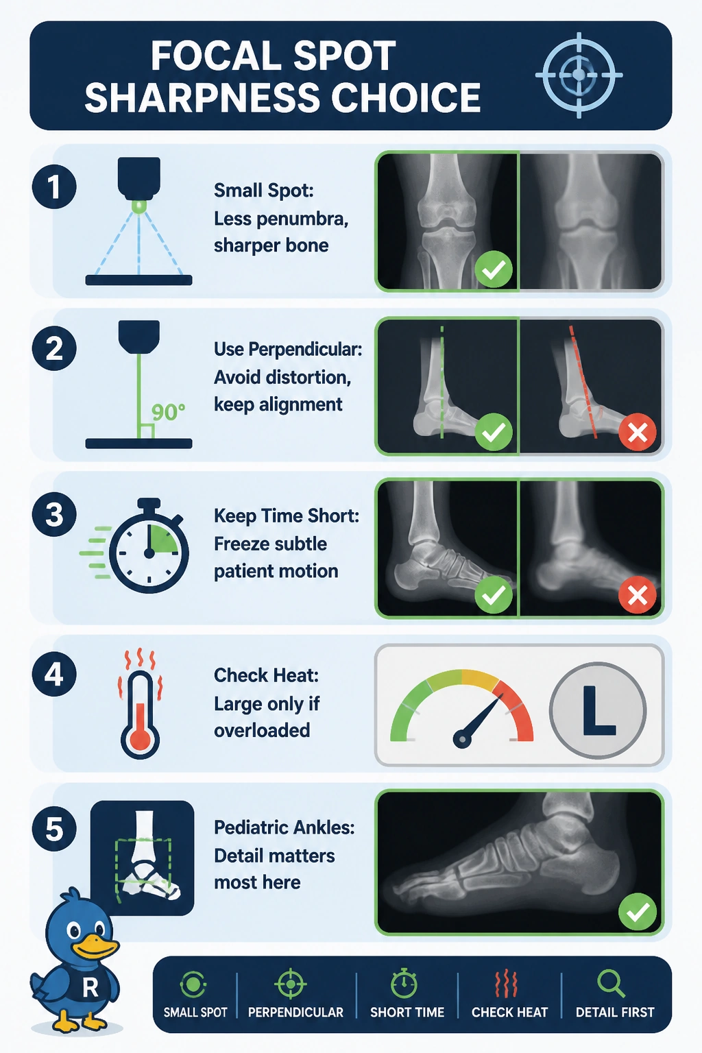
You are squeezing every drop of sharpness here! Tiny anatomy loves a tiny source, so the small focus gives you crisp trabeculae without heat worries.
Full Explanation
For a thin pediatric ankle imaged with ample heat capacity and a young patient who may move, recorded detail is limited mainly by geometric unsharpness and alignment, so the small focal spot is ideal because its smaller effective source reduces penumbra and improves spatial resolution without overheating risk on a high-output tube, the perpendicular central ray preserves true shape and avoids elongation or foreshortening when the foot is already dorsiflexed to 90 degrees, and the short exposure minimizes motion unsharpness while freezing any involuntary movement. The large-focal-spot perpendicular short-exposure technique is routine for thick adult body parts or high-heat-load sequences to distribute anode heat and permit higher mA, but here it needlessly enlarges the penumbra and degrades fine trabecular and physeal detail despite ample cooling. The small-spot angled short-exposure technique is standard for a plantar-flexed ankle to open the tibiotalar joint when dorsiflexion is impossible, but with the foot dorsiflexed angulation introduces shape distortion and degrades alignment without adding coverage. The small-spot perpendicular extended low-mA technique is a valid low-noise strategy to smooth quantum mottle by increasing mAs, but prolonged exposure time increases motion unsharpness and directly opposes the goal of maximal detail. Exam caveat: do not trade increased mAs and reduced noise for detail when the question asks for maximal recorded detail, since noise and sharpness optimize differently. Practical check: confirm small spot selected, central ray perpendicular to the part and receptor, and use the shortest exposure with appropriate immobilization and SID to verify crisp cortical margins.