
A radiographer images a small pediatric hand at 45 kVp and then an obese adult abdomen at 85 kVp. The educator asks why contrast differs and which photon-tissue process dominates in each case. Which interaction explanation correctly links kVp, tissue attenuation, and image contrast?
Select an answer to reveal the explanation.
Short Explanation
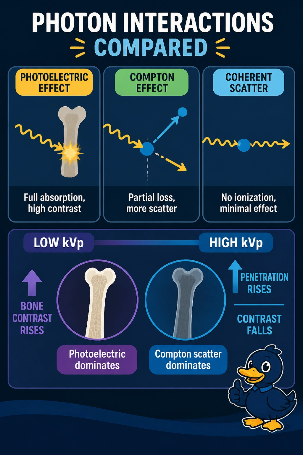
You’re reading contrast like a story of photon choices. Low energy favors full absorption in bone, so photoelectric action gives you that crisp black-and-white pop.
Full Explanation
The correct explanation reflects energy and atomic number dependence of attenuation: photoelectric absorption varies roughly as Z cubed over E cubed, so at 45 kVp calcium in bone with high effective Z absorbs disproportionately and creates high subject contrast in the small hand, while at 85 kVp in the thick adult abdomen photon energies exceed K-edge thresholds and Compton interaction governed by electron density dominates, lowering contrast and producing scatter fog. Attributing low-kVp photoelectric dominance to soft tissue rather than bone fails on tissue dependence because low-Z muscle and fat show little photoelectric enhancement and yield only small density differences. Reversing the energy pattern to place photoelectric effect at high kVp and Compton at low kVp fails on energy dependence because photoelectric probability falls steeply with rising energy while Compton declines slowly and prevails in soft tissue above about 30 keV. Invoking coherent scatter as dominant at high kVp fails on interaction significance because coherent change of direction without ionization contributes only a few percent in the diagnostic range and does not drive contrast. Exam caveat is to separate Z effects, kVp effects, and thickness effects when stems combine anatomy and technique. Practical check is to compare a crisp low-kVp hand with stark bone contrast to a flatter high-kVp abdomen that needs a grid for scatter cleanup, confirming the shift from absorption contrast to Compton transmission.