
An outpatient center images a large adult abdomen supine at 80 kVp without a grid, using 20 mAs with tight collimation. The supervisor adds an 8:1 grid for the next similar patient to reduce scatter while keeping kVp and SID constant. Which grid conversion and collimation adjustment best preserves density while limiting scatter?
Select an answer to reveal the explanation.
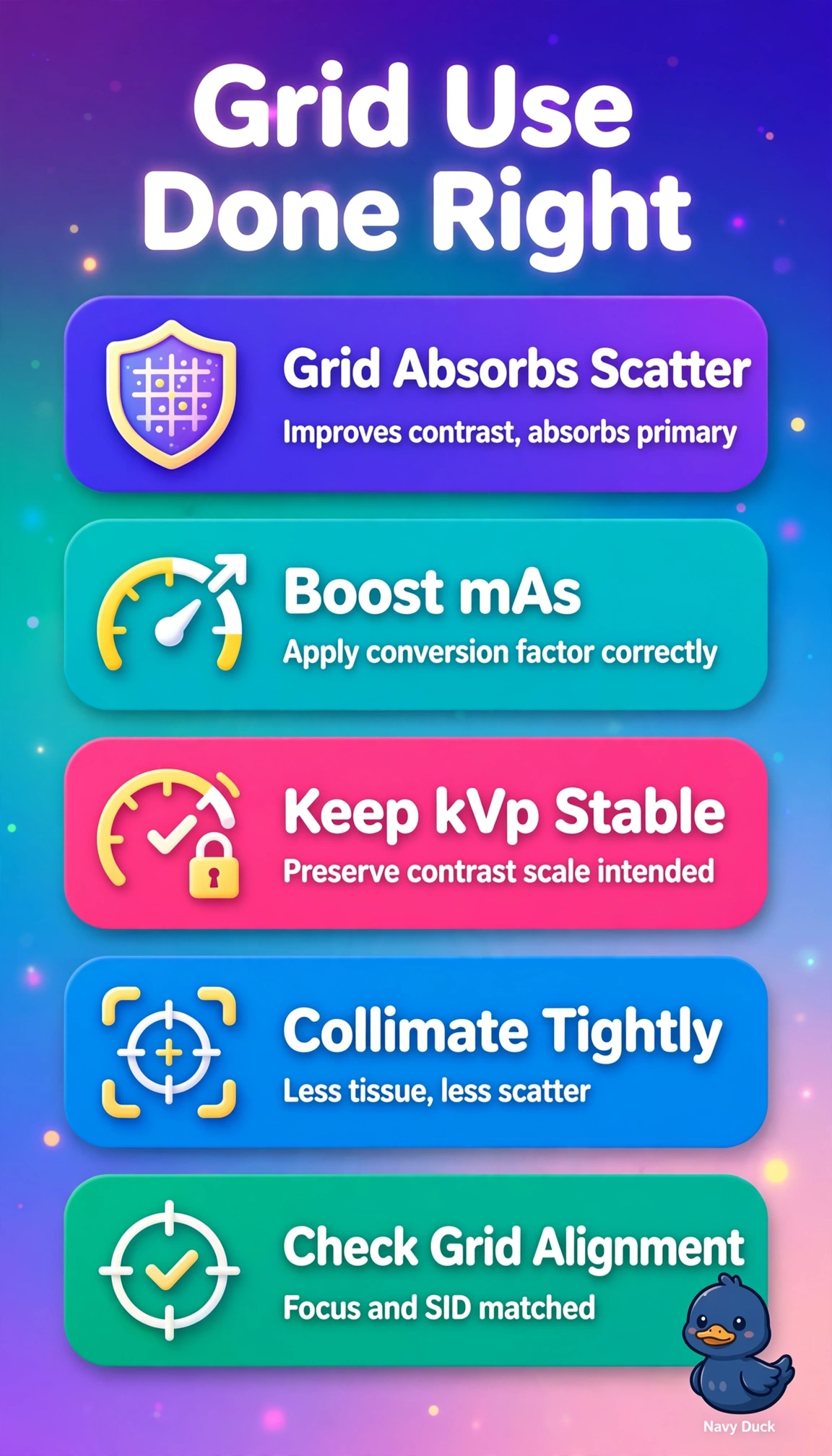
Short Explanation
You nailed the trade-off here — grids clean up scatter but eat primary too. Keeping collimation tight plus boosting mAs by the conversion factor keeps density steady without extra fog.
Full Explanation
Adding an 8:1 grid for a large adult abdomen at 80 kVp removes much scatter but also absorbs primary photons, so density can only be preserved at constant kVp and SID by increasing mAs by the grid conversion factor, about four times for this ratio, while tight collimation to the anatomy further limits scatter production, fog and unnecessary dose and preserves contrast. Widening collimation to ensure full abdominal coverage while correctly raising mAs maintains receptor exposure but defeats the scatter goal because larger field size creates more scatter volume and fog, lowering contrast despite grid cleanup. Raising kVp for penetration while raising mAs and keeping tight collimation is common for thick anatomy, yet it violates the explicit keep-kVp-constant condition and changes the contrast scale, penetration and scatter fraction, so it does not preserve the original image characteristics. Increasing mAs only slightly below the conversion factor with tight collimation reflects sound dose-saving intent, but under-compensation leaves underexposure with low density and increased quantum noise, failing the requirement to preserve density. Exam caveat is to separate grid ratio from conversion factor and avoid applying the 15 percent kVp rule when the stem fixes kVp, focusing instead on mAs scaling and field-size control. Practical check is to confirm tight light-field borders, verify the calculated mAs, then review density and the exposure indicator before releasing the patient.