
A 19-year-old college athlete presents to urgent care after a elbow to the face, with periorbital swelling, double vision on upward gaze, and suspected orbital floor fracture. The provider requests facial bones without CT initially. The patient can sit upright and extend the neck. Which projection and positioning best demonstrates the orbits and sinuses for this suspected blowout injury?
Select an answer to reveal the explanation.
Short Explanation
Double vision when you look upward points you toward an orbital floor injury. With the Waters view, you open the orbits to best demonstrate the suspected fracture.
Full Explanation
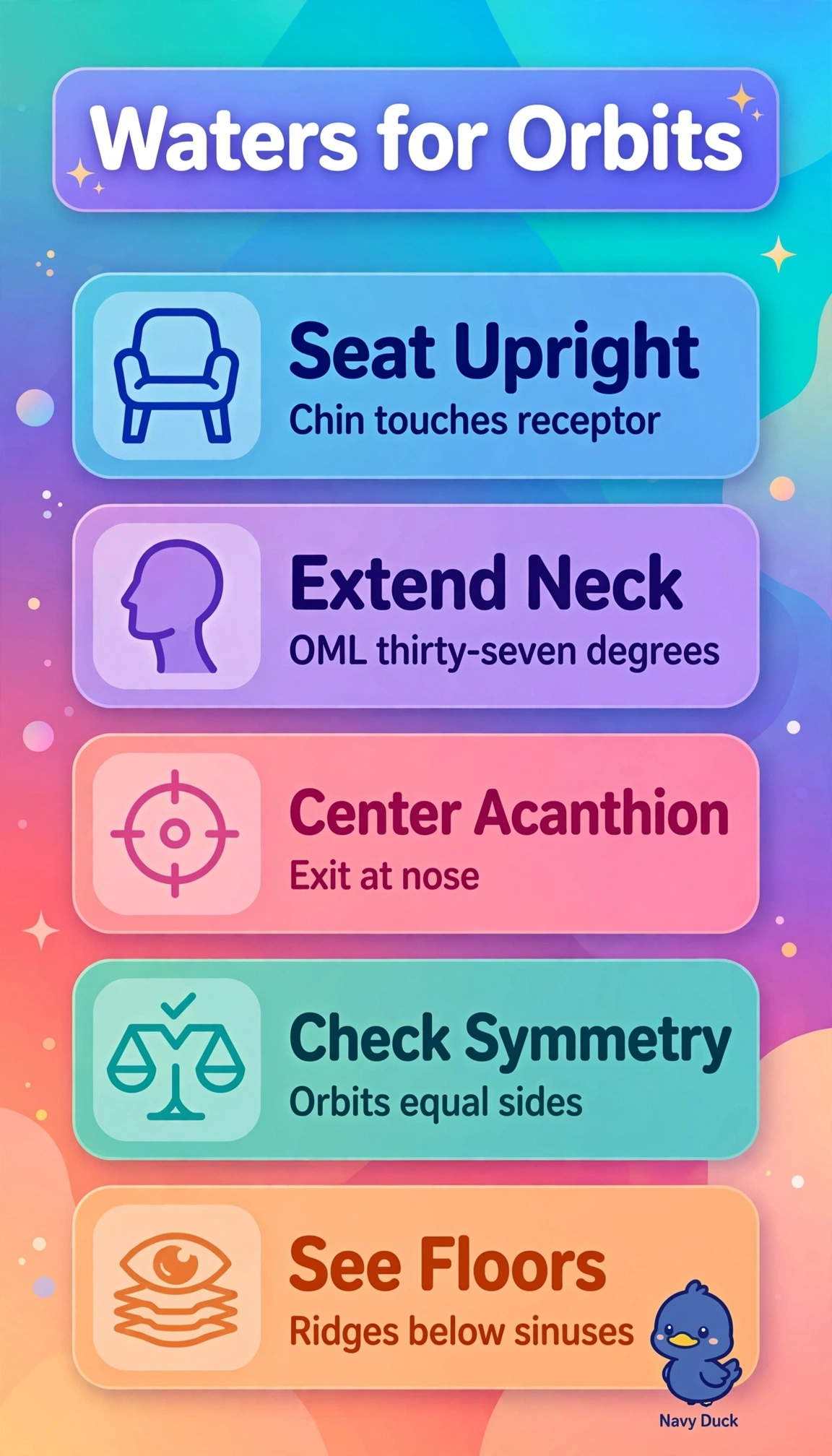
The parietoacanthial Waters projection is the best choice for this suspected blowout injury: position the patient upright with the chin on the receptor and neck extended until the orbitomeatal line forms about 37 degrees with the image receptor; the central ray exits the acanthion. This projects the petrous ridges below the maxillary sinuses and demonstrates the orbital floors, rims, maxillae, and zygomas. A lateral nasal bones projection isolates the nasal bridge in a true lateral view, so it cannot demonstrate the orbital floors or air-fluid levels. A submentovertex projection demonstrates the zygomatic arches and sphenoid sinuses, but superimposes facial structures and increases lens dose without profiling the orbital floors. An AP Caldwell projection, with the forehead and nose on the receptor and the orbitomeatal line perpendicular to it, demonstrates the frontal sinuses, ethmoids, and superior orbital fissures rather than the inferior orbital margins. Exam caveat: associate Waters with orbital floors and Caldwell with frontal and ethmoid sinuses. Practical check: before accepting the image, confirm that the petrous ridges lie just below the sinuses and that the orbits are symmetric, indicating no rotation.