
A 7-year-old referred by pediatrics for short stature arrives with a guardian asking about growth-plate imaging, and the order reads bone age study with no trauma history. The department protocol requires minimal dose and a standard reference for maturity. Which protocol best evaluates skeletal maturity for the bone age study?
Select an answer to reveal the explanation.
Short Explanation
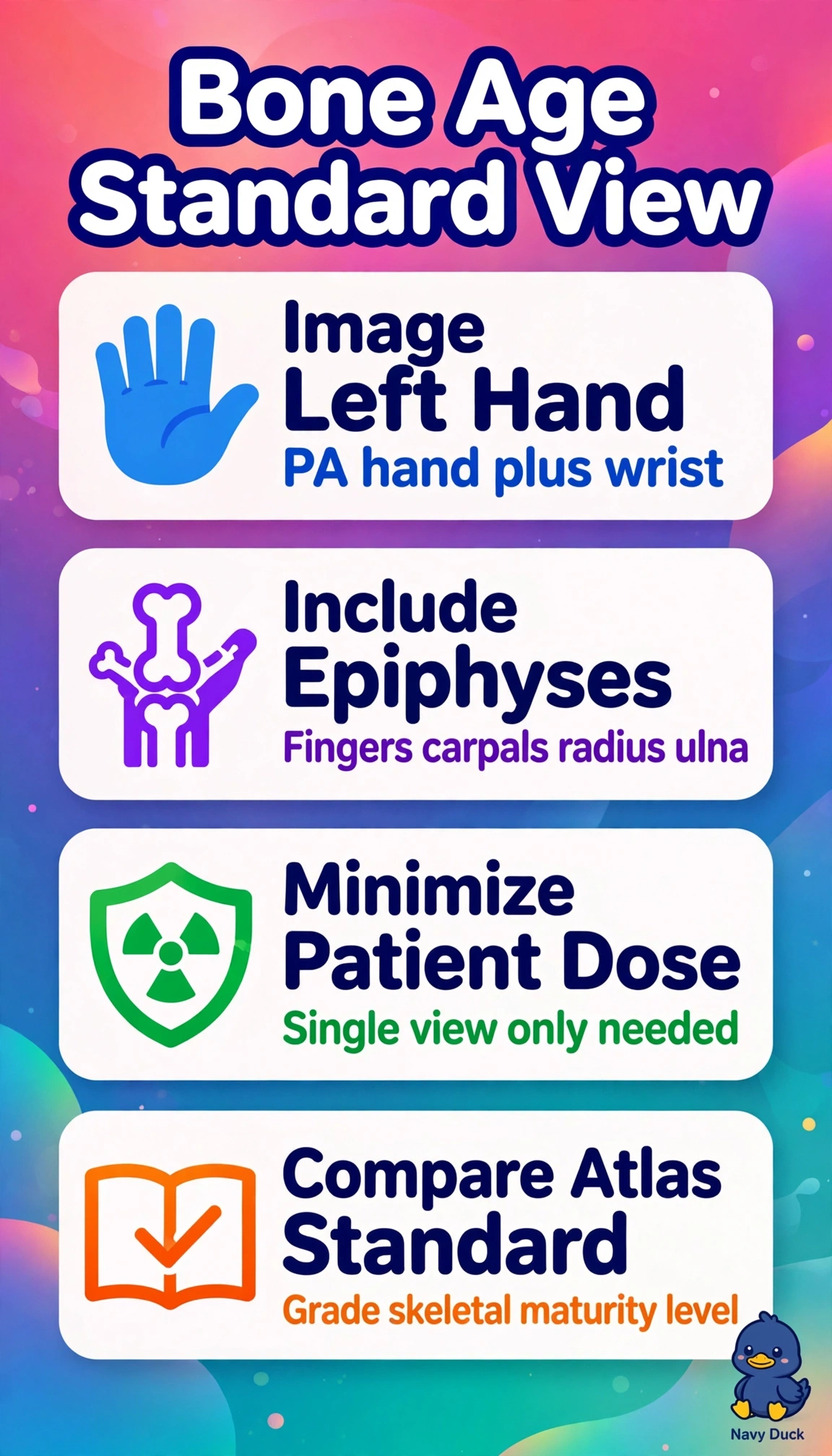
You’re checking one ripening leaf, not the whole tree. A single PA view of the left hand and wrist gives you a clean, low-dose maturity match using a standard atlas.
Full Explanation
A PA view of the left hand and wrist is the accepted bone-age standard because the Greulich-Pyle and Tanner-Whitehouse atlases were validated on that anatomy. It captures many ossification centers in one small, low-dose field, making it suitable for a 7-year-old with short stature and no trauma. A PA view of the right hand and wrist can be used when the left is injured, but without that exception, using the validated left side maintains standardization and comparability. PA and oblique left hand views resemble routine hand-trauma imaging, but the added view increases dose without improving maturity grading. An AP view of the left knee may be used for other assessments, such as leg-length or pubertal staging, but it is not the validated hand-wrist reference for this bone-age study. Exam caveat: do not substitute a pain, injury, or arthritis protocol when the order explicitly requests a bone-age study; the indication determines the anatomy and views. Practical check: verify the order and patient identity, explain the single quick exposure to the guardian and child, place the correct left marker, position the palm prone with fingers extended, and collimate tightly from the distal radius and ulna through the fingertips.