
A busy ICU team requests daily portable chest images on a thin adult using a digital detector with high detective quantum efficiency and wide exposure latitude. Images look acceptable but doses are creeping up and documentation is incomplete. Which grid and detector strategy best minimizes patient exposure while preserving diagnostic quality?
Select an answer to reveal the explanation.
Short Explanation
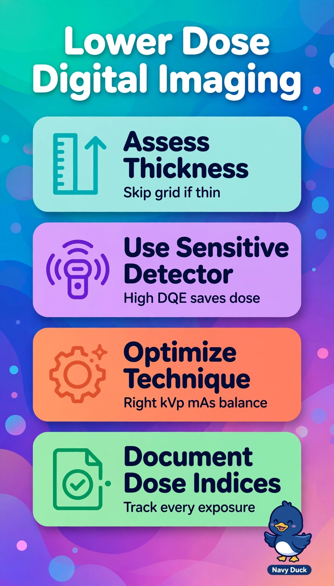
You do not need heavy armor for a light job. Ditching the grid when anatomy is thin cuts scatter cleanup and dose while a sensitive detector keeps quality strong.
Full Explanation
Correct approach works because thin parts generate minimal scatter so removing grid eliminates Bucky factor dose penalty without degrading contrast, high-DQE detector yields higher signal-to-noise per unit exposure allowing mAs reduction, and recording technical factors plus dose indices reveals creep that wide latitude otherwise hides despite acceptable appearance, preserving diagnostic quality for daily portables. Keeping low-ratio grid fails on scatter-grid balance because even low-ratio grids attenuate primary beam and require higher mAs for equal detector dose, unjustified for thin adult chest. Using standard-efficiency portable detector fails on DQE principle because lower efficiency demands greater incident exposure for equivalent noise, increasing cumulative dose from serial exams. Fixing mAs without tracking indices fails on exposure management because digital latitude maintains brightness across overexposure, so undocumented drift continues and incomplete records prevent optimization. Caveat for exams is acceptable look does not confirm correct exposure, and grids are still warranted for thick anatomy where scatter contrast loss outweighs dose. Practical check is verify exposure deviation index against target on each image, confirm technique matches thickness chart, and audit logged factors and indices for upward trend.