
A 14-year-old gymnast has uneven shoulders and a rib hump on forward bend, suggesting idiopathic scoliosis. She can stand erect without support, has no trauma, and the orthopedist needs full-spine curvature measurement with minimal breast dose. Which scoliosis projection protocol best demonstrates curvature while minimizing dose?
Select an answer to reveal the explanation.
Short Explanation
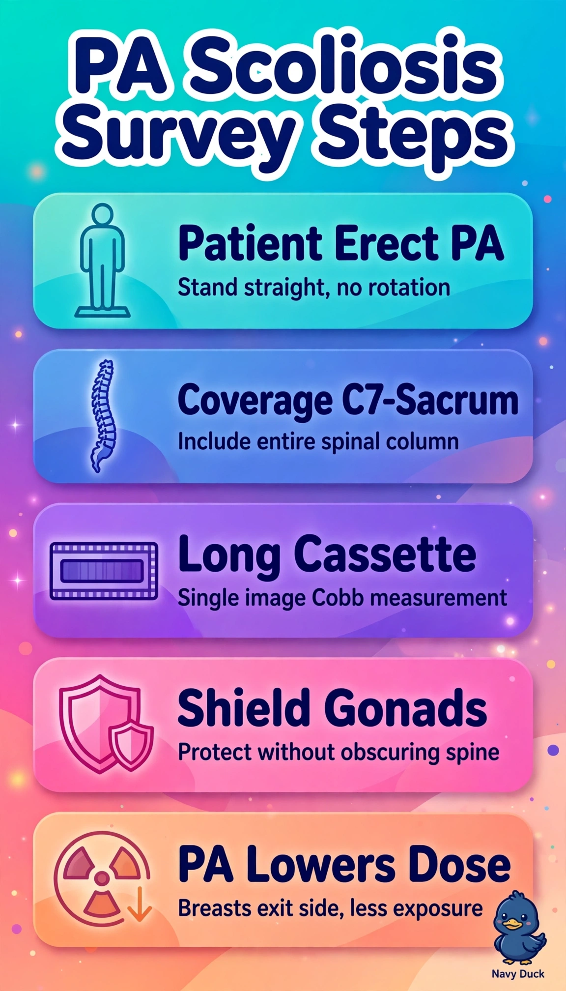
You've got to show the whole curve under gravity while sparing dose. An erect PA long film captures alignment from neck to pelvis with dose-sparing geometry like a shadow falling away from you.
Full Explanation
Scoliosis evaluation requires weight-bearing demonstration of lateral curvature and rotation across the entire spine, typically from C7 through the sacrum on a single long erect image for Cobb angle measurement, and the PA projection is preferred because the incident beam enters posteriorly, placing breast, thyroid, and lens tissue on the exit side where dose is markedly lower while still displaying vertebral bodies and pedicles for curvature assessment. The AP erect full-spine approach demonstrates anatomy and coverage but fails by concept of dose optimization because anterior entry substantially increases radiosensitive breast and thyroid exposure without added diagnostic benefit. The supine spot lumbar approach fails by concept of functional assessment because non-weight-bearing and limited coverage removes gravitational loading and hides compensatory thoracic curves essential to scoliosis classification. The erect bending thoracic approach fails by concept of examination purpose because bending views are supplemental for flexibility, not the initial curvature survey, and limited coverage cannot measure global balance. For the exam, remember erect PA long-spine is the default scoliosis survey unless the question specifies otherwise, and in practice verify C7 to sacrum inclusion, straight posture without rotation, and proper gonadal shielding placement before exposure.