
A patient with a lung lesion that moves with breathing is set up for stereotactic treatment. The department offers daily orthogonal kV imaging, MV portal images of the treatment fields, and CBCT, and the lesion sits away from any fixed bony landmark. Which imaging strategy best verifies the target position for each fraction?
Select an answer to reveal the explanation.
Short Explanation
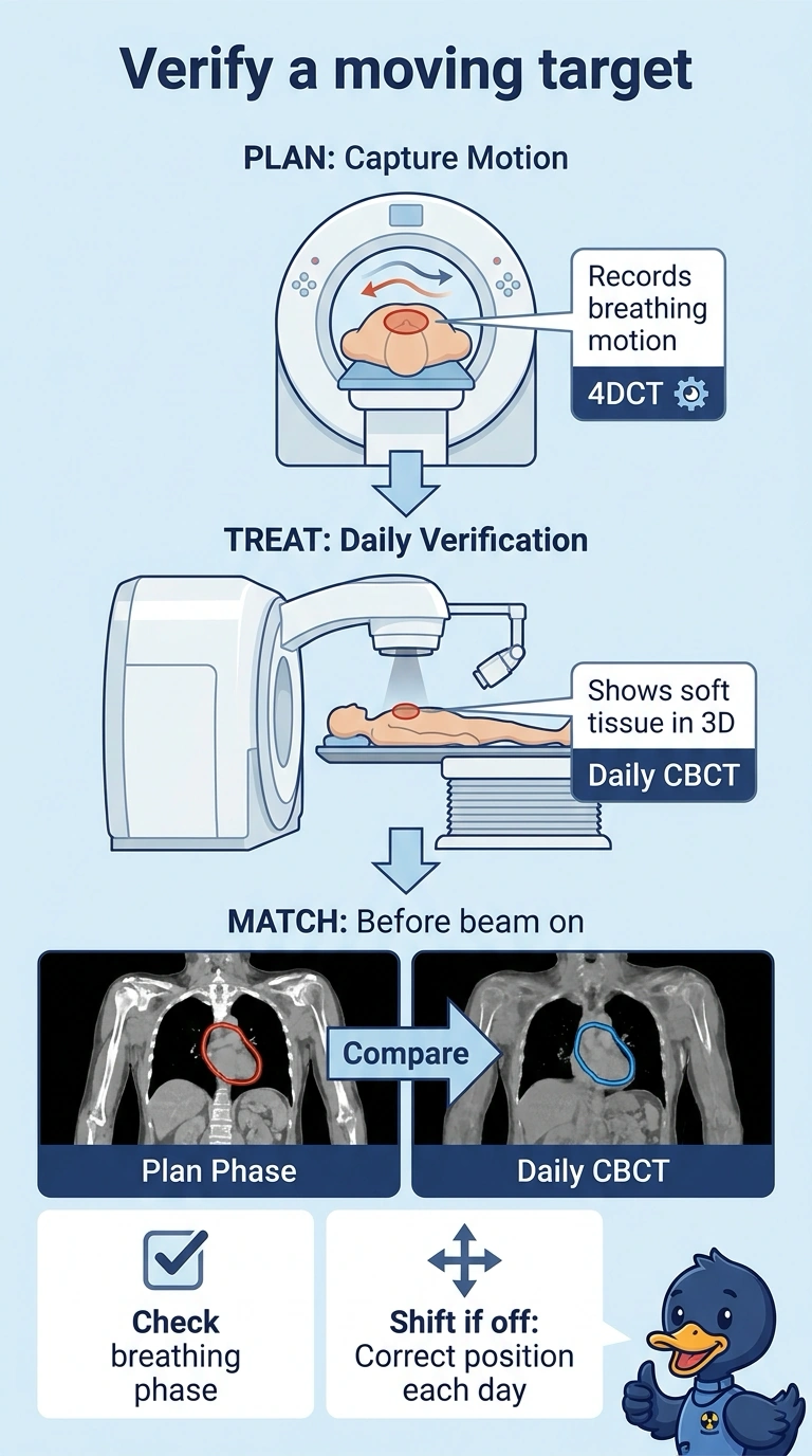
Bony landmarks won't find a lung target that slides with every breath, so you need an image that shows the tissue itself. Daily CBCT paired with a 4D plan gives that check before beam-on each fraction. Seeing the soft tissue on a daily scan is your safety net.
Full Explanation
A target that moves with breathing cannot be located by bone or skin marks, so daily volumetric imaging is what makes the setup dependable. CBCT produces a three-dimensional soft-tissue image in the treatment room, letting you match the target on the planning CT to the target as it sits on the table, and repeating it each fraction catches day-to-day position change. Pairing that verification with a 4DCT-based plan means motion was characterized when the target volume was defined, so the image can be judged against the same breathing phase. Orthogonal kV imaging fails the anatomy requirement because it matches bone at the isocenter and this lesion has no fixed bony neighbor. Planning from a single free-breathing scan fails the motion requirement, because one scan averaged across the breathing cycle leaves no defined phase to match the daily image against. MV portal imaging fails the visualization requirement, because it confirms field geometry and leaf shape rather than where soft tissue sits. Exam caveat: whichever image is used, the match is reviewed and any shift applied before beam-on. Practical check: confirm the daily CBCT is queued in the record, that it is matched to the correct breathing phase, and that shifts are documented.