
A patient who cannot be raised to isocenter is treated at an extended SSD of 115 cm for a parallel-opposed abdominal field; the chart lists an 18 x 20 cm field and a midplane depth of 14 cm. At what distance from the source should the light field be measured to verify the stated size?
Select an answer to reveal the explanation.
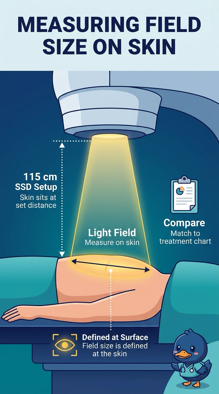
Short Explanation
Your field size and treatment depth are measured on different planes, so they don’t match. With an SSD setup, you measure the light field on the skin at 115 cm from the source—it’s like checking a doorway at the threshold, not at the far wall.
Full Explanation
The correct measurement plane is the patient’s skin at 115 cm from the source. This is an SSD setup, and the stated field size must be checked at the actual treatment surface: beam divergence means the field dimensions there differ from those at another source distance. Measuring at the 100 cm isocenter plane is a standard way to specify or check field dimensions for an SAD treatment, but it does not verify the field size at this patient’s 115 cm SSD. A phantom measurement at 100 cm SSD is also a familiar machine or setup check, yet it substitutes a reference distance for the patient’s actual surface distance and therefore cannot confirm the stated field at treatment. An EPID image can be useful for verifying the radiation field during treatment, but it does not measure the light field requested here; agreement between the radiation field and light field should not simply be assumed. Exam caveat: field dimensions may be quoted at a reference plane such as isocenter, so check the chart’s convention before comparing numbers; that convention does not change the measurement plane required by this question. Practical check: confirm the source-to-skin distance is 115 cm and assess both field edges on the skin with the light field, keeping the patient and beam geometry unchanged.