
A female patient is simulated for treatment of the cervix, and the therapist outlines organs at risk on the planning CT before the plan is scored. Which relationship between the cervical target and the nearby pelvic organs is correct on axial images?
Select an answer to reveal the explanation.
Short Explanation
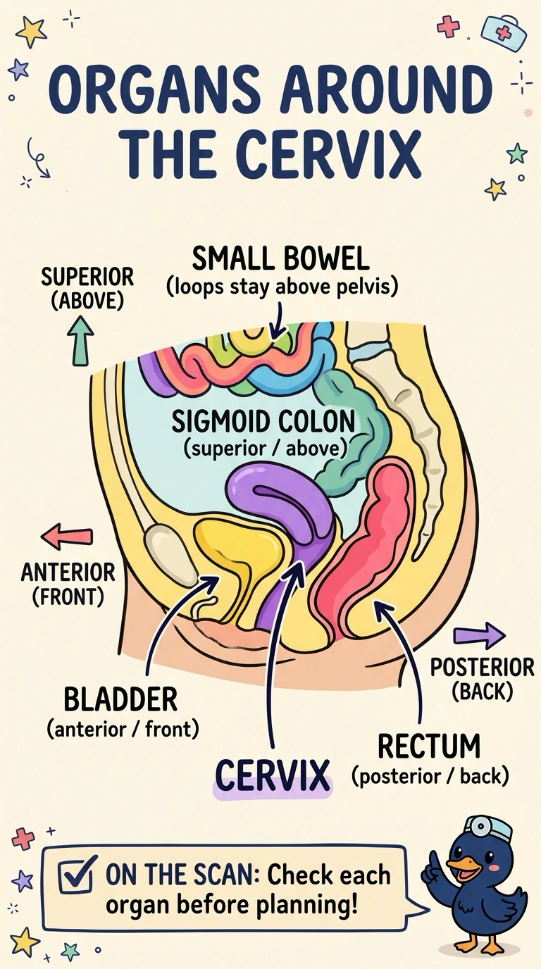
You read the axial slice the way the scanner does, with anterior at the top of the image when the patient lies supine. The cervix sits in the true pelvis, with the bladder cradled in front, the rectum behind and the sigmoid loop above. Keep that order and you'll see why the organs at risk outline themselves.
Full Explanation
On axial images of the true pelvis the bladder sits immediately anterior to the cervix, the rectum lies directly posterior, and the sigmoid colon arches superior to the uterine body, so those relationships define the organs at risk around a cervical target. Swapping anterior and posterior fails because it would place the bladder behind the target and the rectum in front of it, reversing two contours the therapist outlines daily. Putting the sigmoid behind the cervix fails because the rectum, not the sigmoid, occupies that space at this level, while the sigmoid belongs to the bowel above the target. Siting the small bowel inferior to the cervix fails because those loops normally lie above the true pelvis, which is why the bowel bag is edited above rather than below a pelvic target. The exam often tests anatomy through organ-at-risk contours instead of a labelled diagram, so think about what you would outline. Before you approve a plan, scroll the axial stack and identify each organ by its own shape and position rather than by its label alone.