
A patient with a lung lesion is scheduled for stereotactic treatment in three fractions, and the lesion moves noticeably with breathing near the chest wall. The department offers kV cone-beam CT, MV portal imaging, kV orthogonal pairs, and surface-guided optical tracking. Which imaging method best confirms that this patient's lesion sits in the planned position before each fraction?
Select an answer to reveal the explanation.
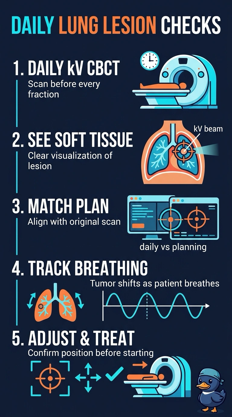
Short Explanation
What you need to see is the lesion itself, and only kV cone-beam CT puts that soft tissue on screen in 3D. The other methods can look perfect while checking just the frame around the target. It's the difference between measuring the frame and checking where the target sits.
Full Explanation
Daily verification for a lesion sitting in aerated lung depends on seeing soft tissue, and kV cone-beam CT is the only listed method that reconstructs a three dimensional image with enough contrast to show the lesion and the lung around it rather than only bone. Because the lesion shifts with breathing, a volumetric match also lets you compare the whole treated volume against the planning scan and decide whether to adjust before the beam starts. MV portal imaging relies on absorption differences that make bone stand out while lung soft tissue washes out, so a clean bony match can still hide a target that is off position. Orthogonal kV pairs are quick and low in dose, but two planar images cannot show where the lesion sits in three dimensions when motion has blurred its edges. Surface guidance tracks skin markers and chest wall contour, which reflects external motion only and says nothing about the internal target. The exam caveat is that cone-beam CT is also used for bony matching, so read the stem for the tissue that actually has to be verified. As a practical check, ask whether your image would let you see the target itself before the beam is turned on.