
A patient with a large anterior-posterior separation needs a photon field set at SSD 118 cm rather than the usual 100 cm, keeping the same field size at the skin. For the geometric inverse-square correction to the primary fluence at 7 cm depth, which factor gives the dose per monitor unit at the extended SSD relative to the usual SSD?
Select an answer to reveal the explanation.
Short Explanation
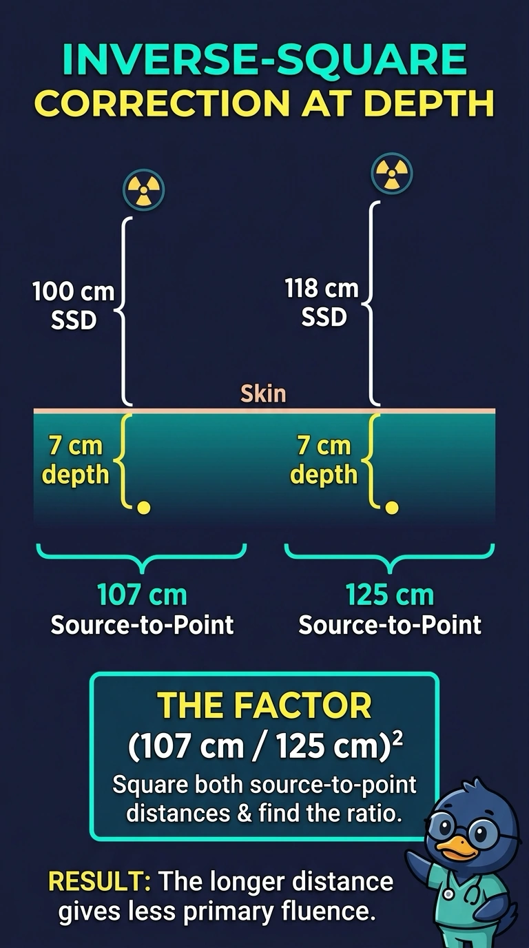
At 7 cm depth, your source-to-point distances are 107 cm at the usual SSD and 125 cm at the extended SSD. Like a beam spreading as it travels, primary fluence follows the inverse-square rule, so the dose-per-monitor-unit factor is (107/125)², or about 0.73.
Full Explanation
For the geometric inverse-square component of primary fluence, use the source-to-point distance, not merely the distance from the source to the skin. At 7 cm depth, the usual-SSD setup has a source-to-point distance of 100 + 7 = 107 cm, while the extended-SSD setup has 118 + 7 = 125 cm. Therefore, the dose-per-monitor-unit ratio at extended SSD relative to usual SSD is (107 cm/125 cm)², approximately 0.73: the greater source-to-point distance reduces primary fluence. The SSD-only inverse-square approach, (100/118)², fails because it omits the point’s depth. A percentage-depth-dose ratio alone describes depth-dose behavior, not this geometric inverse-square correction. A surface-field-area ratio alone describes field-size geometry and likewise does not give the source-to-point distance correction. Exam caveat: this is only the geometric component; a complete clinical dose calculation may also require appropriate depth-dose, output, and field-size corrections. Practical check: add depth to each SSD before forming the ratio, and confirm the extended-SSD factor is less than 1.