
A hemithorax field for a large patient is too wide for the department's standard 100 cm SSD, so the physicist plans treatment at 120 cm SSD and recalculates before the first fraction. Compared with the 100 cm setup, how do beam output and percentage depth dose change at 120 cm SSD?
Select an answer to reveal the explanation.
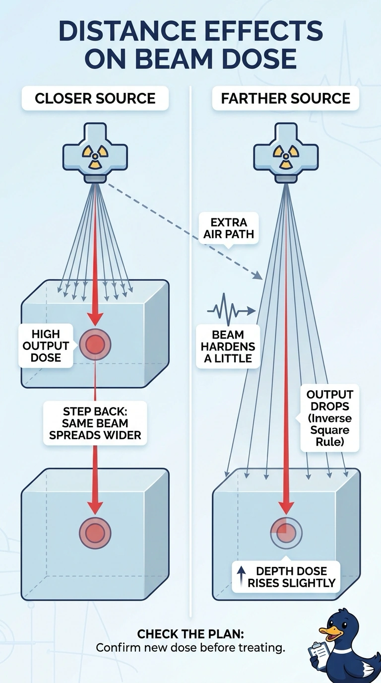
Short Explanation
Step the source back and the same beam spreads over a wider area, so your output follows the inverse square law down by the distance ratio squared. The extra air path also hardens the beam a touch, and that's why your depth dose rises slightly at the same depth. It works like a flashlight dimming as you back away from the wall.
Full Explanation
When the source-to-surface distance grows, the photon fluence reaching the skin is shared across an area that scales with the square of that distance, so output follows the old distance divided by the new distance, squared, and at 120 cm that reduction has to be made up with additional monitor units. The depth-dose profile also shifts, because the longer air path filters out more low-energy photons, hardening the beam before it enters the patient, and a harder beam penetrates deeper, so percentage depth dose rises slightly at any given depth. Describing the beam as softening in air reverses the filtration effect, since preferential absorption of low energies is exactly what hardening means. Claiming depth dose stays the same ignores that percentage depth dose depends on source-to-surface distance as well as depth and field size. Calling the drop slight underestimates the geometric divergence of a near-point source. Exam caveat: expect a squared distance factor applied to monitor units. Practical check: confirm the plan's stated SSD and compare the recalculated monitor units with the published output factor for that distance.