
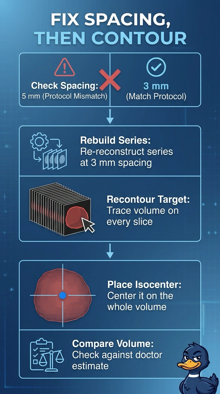
A pelvis planning CT was reconstructed at 5 mm spacing instead of the 3 mm spacing required by the simulation protocol, so the contoured target volume differs from the physician's estimate, and the isocenter sits at the center of the largest axial slice. Which corrections must be completed before the plan is calculated?
Select an answer to reveal the explanation.
Short Explanation
Slice spacing decides how much detail your contours can really capture, so rebuild first and contour second. Placing the isocenter on one fat slice is a flat-screen shortcut in a three-dimensional job. You finish the reconstruction, recontour, then center the isocenter on the whole volume.
Full Explanation
Reconstruction spacing controls how finely anatomy is sampled, so a 5 mm series misses contour detail that a 3 mm series captures, and rebuilding from stored raw data at the protocol spacing lets the contoured volume agree with the physician's estimate before any calculation runs; the isocenter then belongs at the center of the entire contoured volume rather than the middle of one large slice, because beams are designed around a three-dimensional target. The sharper-kernel approach fails because display filtering changes apparent edge contrast without restoring slices that were never acquired. The interpolation approach fails because added in-between slices are mathematically created, producing a smoother-looking volume rather than new anatomic information. The keep-the-slice-center approach fails because it preserves a two-dimensional shortcut that can leave the true volume center off axis. An exam caveat is that slice thickness protocols differ by treatment site and institution, so follow the local protocol rather than a memorized number. A practical check is to confirm spacing, volume, and isocenter on all three planes before the plan leaves the department.