
A physician contours a lung lesion on the simulation CT, and the record must show each target volume as a separate structure for review. The plan has to cover microscopic spread beyond the visible lesion and absorb day-to-day setup variation. Which contour sequence correctly defines the volumes used for the dose plan?
Select an answer to reveal the explanation.
Short Explanation
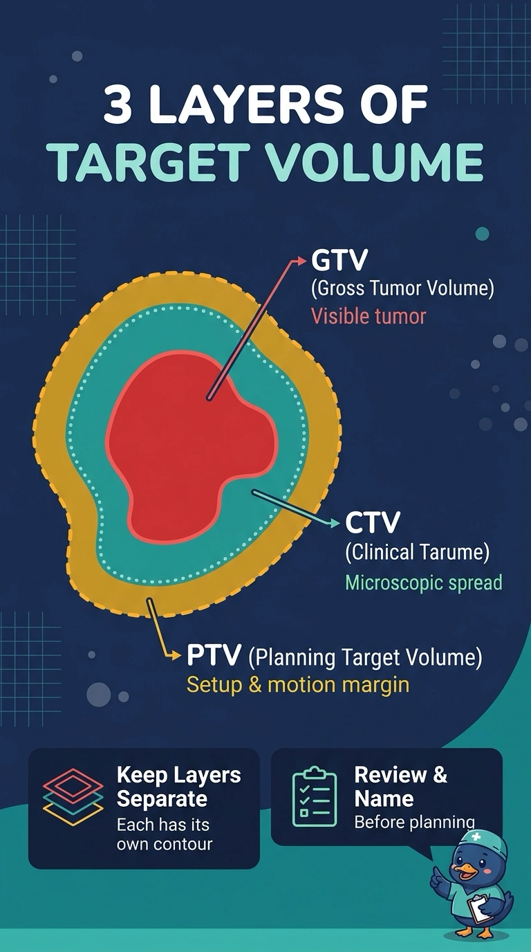
They work like layers of a safety net, and each layer answers a different question. It's easy to blend them, but contour what you see, define the clinical target volume for microscopic spread, then add setup room with a planning target volume.
Full Explanation
The gross tumor volume captures disease that is clinically or imaging-detectable, the clinical target volume includes regions where microscopic disease is plausible, and the planning target volume accounts for geometric uncertainties such as setup and internal motion. Building them in that order keeps each uncertainty identifiable and the record auditable, because a reviewer can distinguish the clinical extent from the margin for positioning and motion. Adding setup margin straight onto the visible lesion fails the requirement for a documented intermediate structure, so microscopic spread outside the visible mass is not separately contoured or reviewed. Stopping at the clinical target volume and calling it final fails the requirement to account for day-to-day setup variation. Blending everything into one structure fails the requirement that each volume appear separately, since a reviewer cannot tell which expansion covers spread and which covers positioning. Exam caveat: margin sizes vary by site, motion, and department protocol, so learn the definitions rather than memorizing a number. Practical check: open the structure set and confirm that each target layer exists, is named, and can be edited on its own before the plan is reviewed.