
A patient with a lung tumor that shifts with breathing completed a 4D CT simulation, and fiducial markers are unavailable. Each day the team must confirm the soft-tissue position of the moving target before treatment. Which imaging approach best satisfies this daily localization requirement?
Select an answer to reveal the explanation.
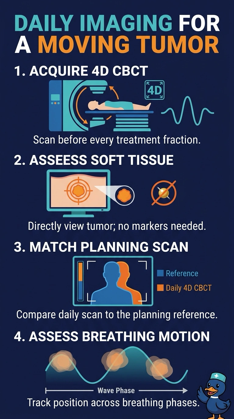
Short Explanation
Because the tumor moves as you breathe, you can acquire respiratory-correlated 4D cone-beam CT before each treatment to track it through the breathing cycle without fiducials. It’s like checking a moving target at several moments, helping you confirm its soft-tissue position before treatment.
Full Explanation
Respiratory-correlated 4D cone-beam CT acquires images in breathing phases, helping the team compare the moving target and surrounding anatomy with the 4D planning CT before each fraction. Tumor visibility on cone-beam CT varies with tumor size, location, and image quality, so the team must confirm that the target is adequately visible. Megavoltage portal imaging has poor soft-tissue contrast, and bony anatomy alone does not confirm the tumor’s position relative to the bones. A single kilovoltage scout with skin marks does not provide a daily three-dimensional check of internal position or respiratory motion. Fluoroscopic tracking of an implanted marker is not an option here because fiducials are unavailable. Image guidance can help manage setup error but does not by itself eliminate planning target volume margins, which also account for motion and other uncertainties. Confirm the imaging mode, dose, and field of view for each fraction and compare the daily image with the reference CT before treating.