
A pathology report on a resected mass describes malignant cells of epithelial origin arranged in solid sheets with no gland formation, marked variation in nuclear size, and mitotic figures in most high-power fields. The oncology team is completing the diagnosis summary for the treatment plan. Which histopathologic type and grade should be recorded?
Select an answer to reveal the explanation.
Short Explanation
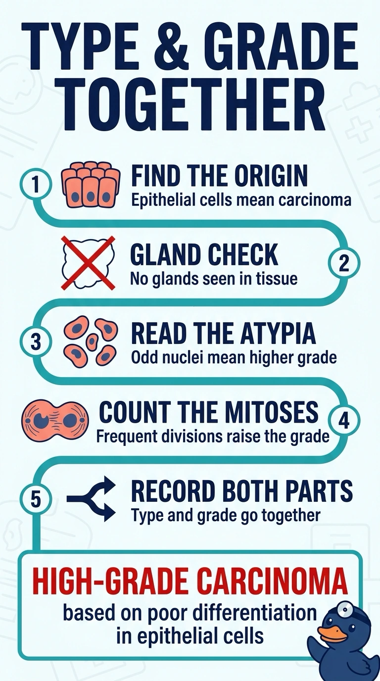
You kept the two clues separate, because histologic type follows where the cells started while grade follows how abnormal they look. The origin clue points to epithelial cells, and the atypia clues point to a high grade. Type's the family tree; grade's how far the cells wandered from it.
Full Explanation
The correct diagnosis is high-grade carcinoma: epithelial origin establishes carcinoma, while solid sheets without gland formation indicate a non-gland-forming pattern, and marked nuclear pleomorphism with mitoses in most high-power fields supports poor differentiation and high grade. A high-grade squamous cell carcinoma is a plausible epithelial malignancy, but the report provides no squamous differentiation, so this specific subtype cannot be assigned from the stated findings. A high-grade neuroendocrine carcinoma can also be highly mitotic and markedly atypical, but those features establish aggressiveness, not neuroendocrine lineage; characteristic morphology or confirmatory immunohistochemistry is not reported. A high-grade adenocarcinoma fits the epithelial origin and aggressive cytology, but adenocarcinoma implies glandular differentiation, directly at odds with the reported absence of gland formation. Exam caveat: absence of glands alone does not identify a specific non-gland-forming subtype; avoid overclassification when lineage-defining features are not provided. Practical check: review the complete microscopic description and, when the differential warrants it, correlate with appropriate immunohistochemical findings before adding a specific carcinoma subtype to the treatment summary.