
A patient with a peripheral lung lesion that moves with breathing is being set up for stereotactic treatment on a linac. The team wants a volumetric check of the tumor's soft-tissue position immediately before beam-on every fraction, with imaging dose kept as low as reasonably achievable. Which imaging modality best meets that daily localization requirement?
Select an answer to reveal the explanation.
Short Explanation
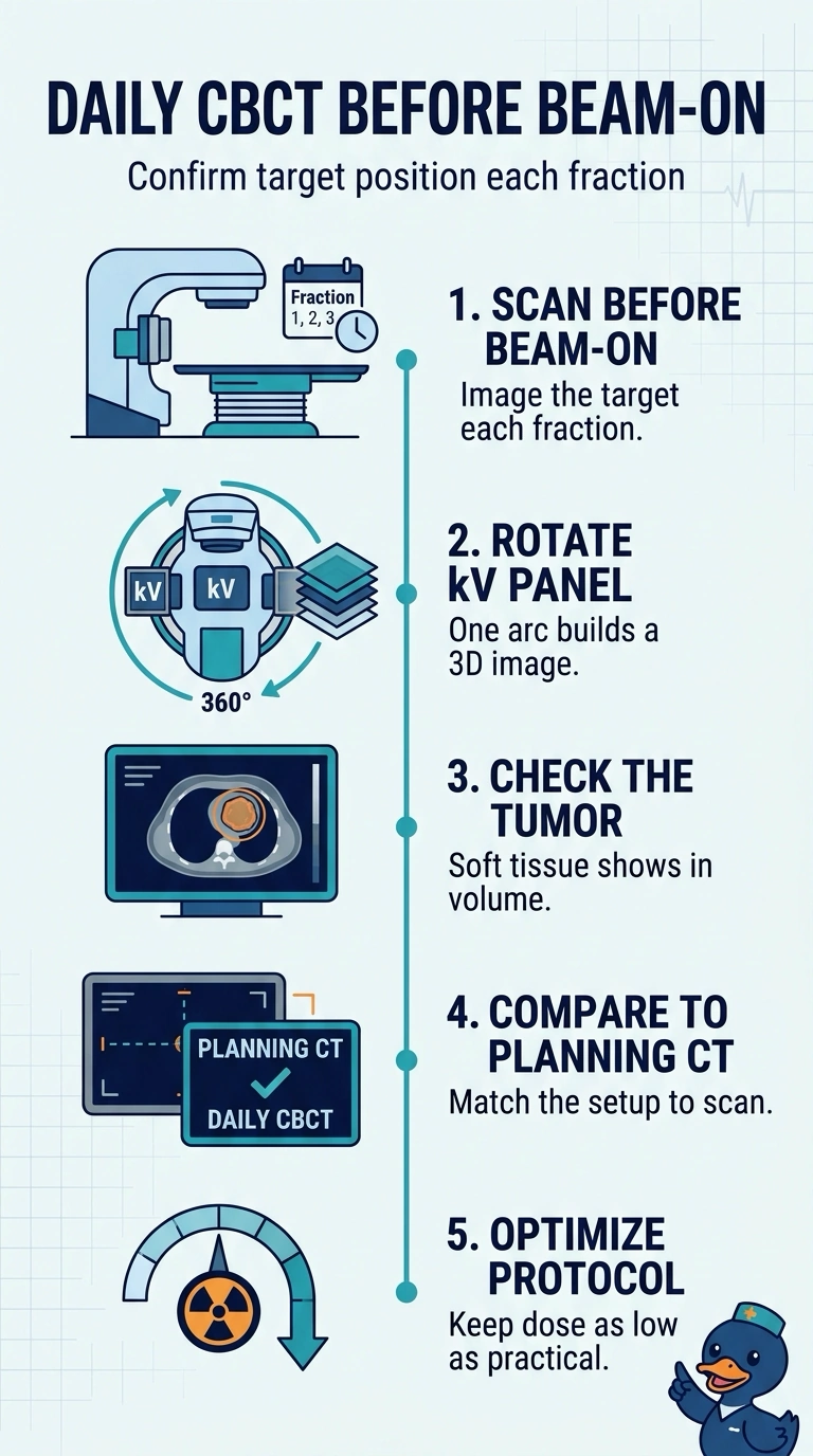
A flat image can line up the bones and still miss where the tumor sits, so you need a volumetric check right before beam-on. Orthogonal kV pictures are quick and can be low dose, but they do not provide a volumetric soft-tissue check. The cone-beam CT shows the soft-tissue target position for that fraction.
Full Explanation
Volumetric guidance right before treatment works because a cone-beam CT rotates an x-ray source and detector around the patient to reconstruct a three-dimensional dataset, helping show where the soft-tissue target sits. Since a lung lesion shifts with breathing, checking it immediately before treatment can reveal day-to-day setup differences that a planar check may not show. The imaging protocol should be optimized to keep imaging dose as low as reasonably achievable. Planar kV verification does not meet the volumetric requirement, and may not show the target clearly without suitable visible landmarks. Weekly verification fails the every-fraction requirement. Megavoltage portal imaging can verify treatment-field geometry, but generally provides less soft-tissue contrast than kV imaging. Exam caveat: imaging choices are matched to target visibility, motion and how often you look, so a method covering only two of those three is rarely the best answer. Practical check: before you commit, confirm the image is volumetric, taken before beam-on, and repeated every fraction.