
A patient with a 3 cm tumor in the right lower lobe is simulated for stereotactic treatment, and fluoroscopy shows the lesion moving almost 2 cm with quiet breathing. The department can offer either a standard helical scan or a gated study, and the physicist needs the motion extent documented before contours are drawn. Which CT simulation acquisition best records the tumor's motion range for planning?
Select an answer to reveal the explanation.
Short Explanation
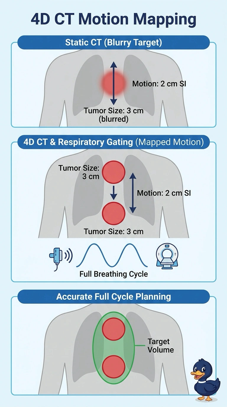
Respiratory motion deserves measurement rather than guesswork, since one snapshot hides it. A 4D CT tags each image with the breathing phase, like a flip book showing the whole trip instead of one frame. You'll use that travel range to build the internal target volume.
Full Explanation
A respiratory-gated 4D CT acquires images continuously while the scanner records the patient's breathing signal, then sorts those images into phases so you can see where the tumor sits at full inspiration, full expiration and every point between. The outer envelope of those positions gives the motion amplitude the physicist uses to build the internal target volume, which is exactly what the request for documented motion extent means. The breath-hold helical approach freezes the lesion at a single phase and therefore never characterizes its travel, leaving any margin built afterward to assumption. The free-breathing scan with a uniform setup margin treats motion as an unknown and pads the target symmetrically, which tends to over-irradiate normal lung while still missing the true excursion. The single deep-inspiration hold delivers crisp images of one extreme but records nothing about the rest of the cycle. An exam caveat is that phase sorting breaks down with irregular or shallow breathing, so artifacts can mimic tumor extent. As a practical check, compare the phase images with fluoroscopy or a prior scan before contours are finalized.