
During a pelvic simulation, the physician needs the loops of small bowel sitting above the bladder to stand out clearly on the axial CT slices so they can be separated from the target. The patient drinks comfortably and the department has cleared them for contrast. Which contrast approach best makes those bowel loops visible for contouring?
Select an answer to reveal the explanation.
Short Explanation
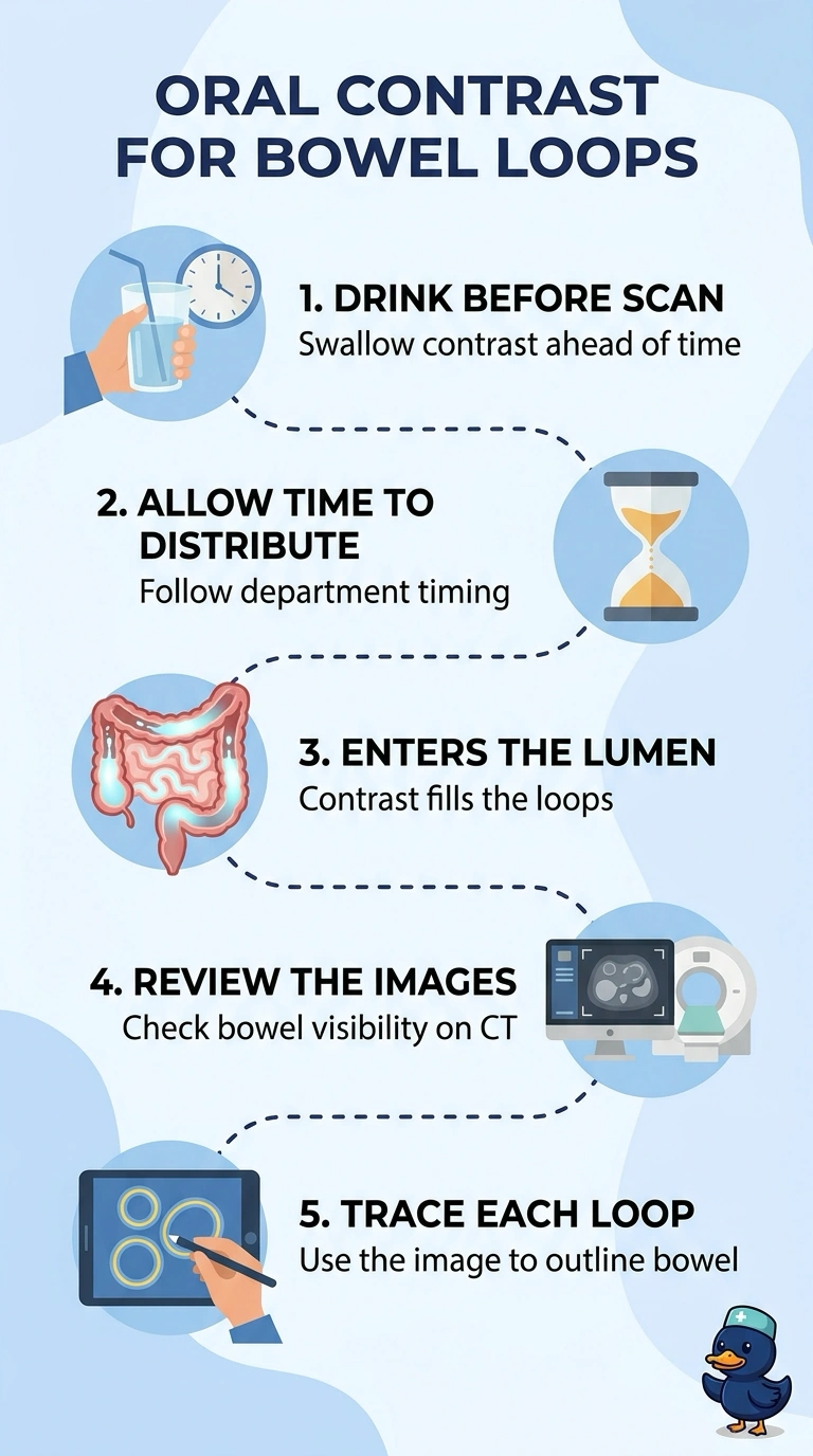
Give oral contrast before the scan so it fills the bowel lumen and helps you see the loops clearly for contouring. It’s like adding colored water to a hose—the contrast highlights the inside, not just the outside.
Full Explanation
Oral contrast is swallowed before the scan and travels into the bowel lumen, where it can increase attenuation and make bowel contents appear bright on axial slices. This may help distinguish small bowel loops sitting above the bladder from the target volume; the visibility and distribution depend on timing and department protocol. Intravenous contrast can enhance bowel walls and vessels, but it does not fill the bowel lumen. Scanning without contrast may still allow bowel identification, but relying on fat planes alone may not make the loops stand out clearly. Rectal contrast is intended to opacify the rectum and distal large bowel and ordinarily does not fill the small bowel loops sitting above the bladder. The contrast route, type, timing, and amount should follow department protocol.