
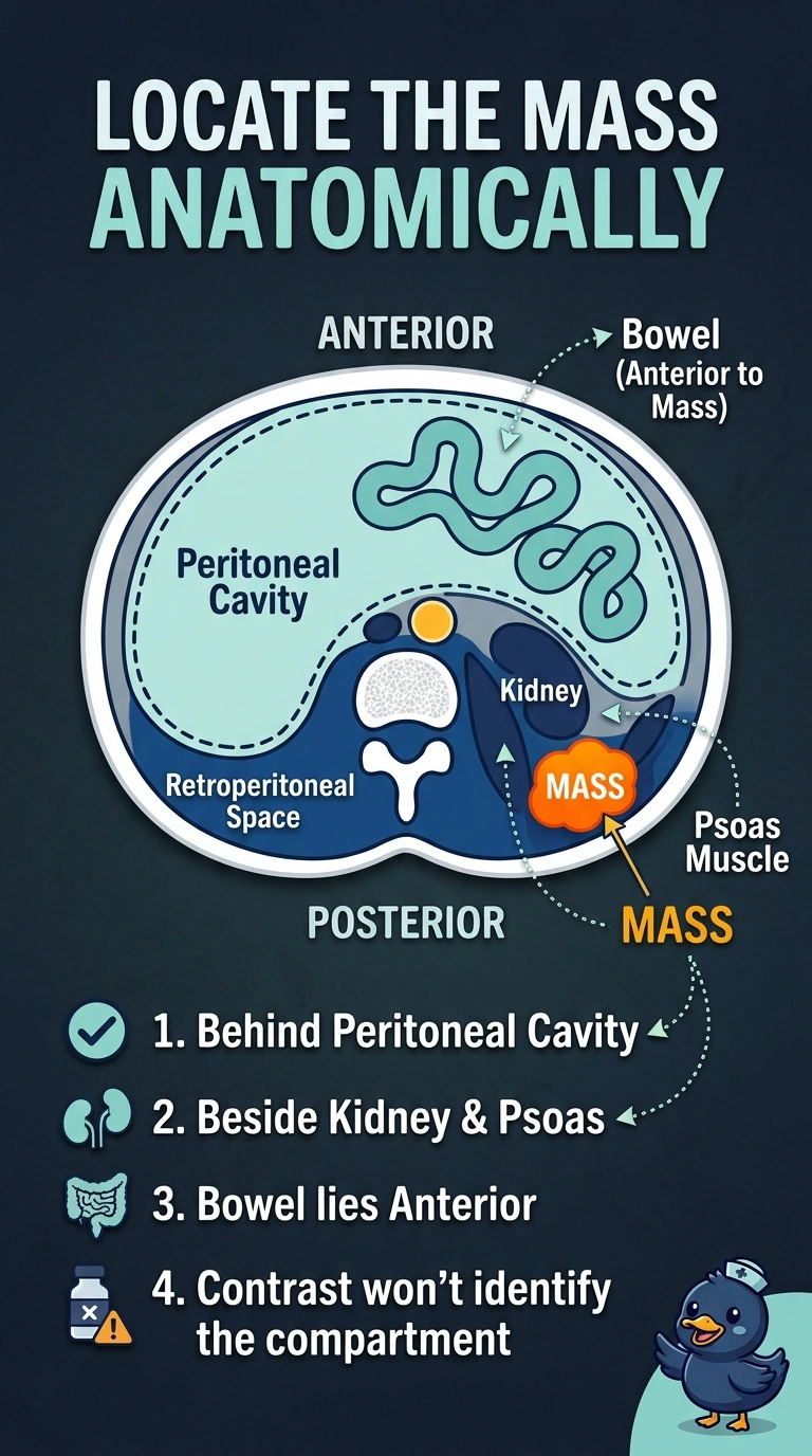
During a simulation CT of the upper abdomen, images are acquired with IV contrast in the portal venous phase. A 3 cm mass lies posterior to the peritoneal cavity, adjacent to the right kidney and psoas muscle, while nearby bowel loops lie anterior to it. Which localization of the mass best fits these anatomic relationships?
Select an answer to reveal the explanation.
Short Explanation
Your mass is retroperitoneal because it sits behind the peritoneal cavity, beside the kidney and psoas, while the bowel lies in front—like a room behind the main living area. The contrast phase helps show the mass, but it doesn’t determine which anatomic compartment it’s in.
Full Explanation
The stated landmarks support a retroperitoneal location: the mass is posterior to the peritoneal cavity and lies beside the kidney and psoas, while bowel loops are anterior to it. These relationships place it behind the peritoneal contents; portal venous contrast timing does not determine the compartment. An intraperitoneal mass may abut bowel, but proximity alone does not place it among the mobile viscera, especially when the bowel is described as anterior to the mass. A renal cortical lesion can lie beside the psoas and enhance with IV contrast, but adjacency to the kidney does not establish origin in renal parenchyma; the description identifies the mass as adjacent, not arising from, the kidney. A psoas-origin tumor would also be retroperitoneal, but contact with the muscle does not prove that the mass originates within it. The exam caveat is that “retroperitoneal” is a broad compartment label; assigning a more specific space or organ of origin requires additional boundaries or evidence of continuity. As a practical check, trace the peritoneal cavity and bowel on sequential axial or multiplanar images, then assess whether the mass is separate from the renal parenchyma and psoas rather than inferring its origin from simple contact.