
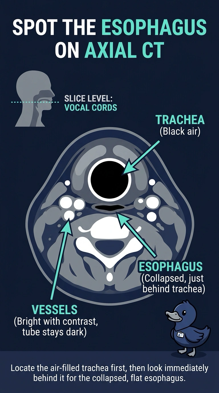
A patient with a T2 glottic laryngeal cancer is simulated on a planning CT with IV contrast, so vessels enhance while the aerodigestive tract stays dark. On an axial slice at the level of the vocal cords you must identify the tube lying directly behind the tracheal air column before labeling the structures on the image. Which structure occupies that position, based on its relationship to the trachea?
Select an answer to reveal the explanation.
Short Explanation
Position is your best clue when soft tissues all look the same shade of gray. Read each slice from the feet up, and you'll see the tube sitting in the trachea's shadow is the esophagus. Contrast brightens the vessels around it, which is why that dark tube stands out so clearly.
Full Explanation
On an axial planning CT you read the slice as if you were standing at the patient's feet, so position against fixed landmarks does the identifying work for you when adjacent soft tissues look alike. The trachea is unmistakable because its air column is black, and the soft-tissue tube tucked directly behind it is the collapsed esophagus lying against the front of the vertebral column at that level. That relationship is what lets you identify the structure confidently while you review the planning images. The aorta fails as an answer because the thoracic aorta runs through the chest to the left of the vertebral column and never sits behind the trachea at the vocal cord level. The thyroid fails because its two enhancing lobes wrap the front and sides of the airway, which places it anterior to the trachea. The spinal cord fails because it is enclosed within the bony spinal canal and never occupies the trachea's posterior shadow. Exam caveat: slice levels drift with body habitus, so confirm the true level using cartilage landmarks instead of counting slices from the top. Practical check: trace the tracheal air column downward, put your cursor on the tissue directly behind it, then confirm that the tube continues into the mediastinum.