
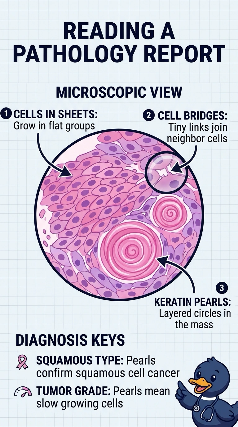
A lung biopsy report describes malignant cells arranged in sheets with intercellular bridges and keratin pearls, and the physician wants the classification entered before planning begins. The team is deciding how the tumor should be labeled. How should this tumor be classified by type and grade?
Select an answer to reveal the explanation.
Short Explanation
When you see keratin pearls and intercellular bridges, they’re like clear signposts pointing to squamous differentiation, so you classify the tumor as squamous cell carcinoma. Because those features are neatly formed, you grade it as well-differentiated.
Full Explanation
Histopathologic classification follows the structures a tumor reproduces from its tissue of origin, and keratin pearls with intercellular bridges are the classic squamous signature, so the lesion is a squamous cell carcinoma; when those features are readily visible the tumor is also graded as well differentiated, meaning it still resembles its parent tissue. Calling it an adenocarcinoma rests on gland or mucin formation, which is not described, so glandular reasoning misidentifies the cell type. Calling it poorly differentiated reverses the finding, because sheets with no recognizable architecture would suggest a high-grade lesion while orderly pearls show retained maturation. Assigning a small cell neuroendocrine type depends on finely granular chromatin, nuclear molding, and crush artifact rather than keratinization, so that pattern does not match what was reported. Be aware that type and grade are separate systems and that grading schemes differ in tier count, so exams usually ask for the defining feature rather than a numeric grade. A practical check: link each descriptive term in the report to the specific structure it names before entering the classification.