
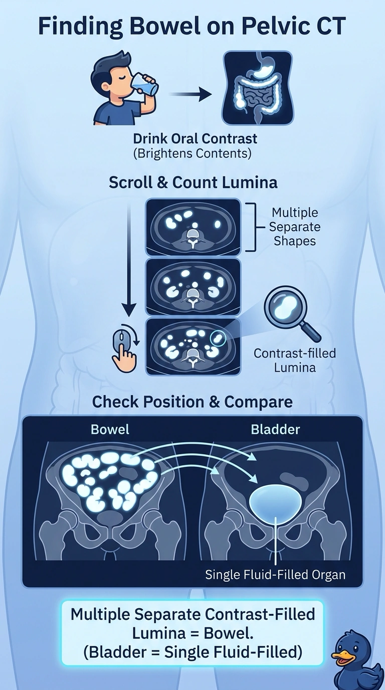
A patient drinks oral contrast before a pelvic CT simulation, and axial images show bright rounded structures low in the pelvis. The therapist must confirm these are bowel loops before the physician contours the target. Which sectional anatomy finding identifies them as bowel?
Select an answer to reveal the explanation.
Short Explanation
When you see several separate contrast-filled lumina, you’re looking at bowel—like a cluster of little pockets, not one large reservoir. Your bladder appears as a single fluid-filled space, so check neighboring slices to follow the structures.
Full Explanation
The correct finding is the presence of multiple separate contrast-filled lumina: oral contrast travels through the gastrointestinal tract, so distinct opacified bowel segments can appear as separate rounded structures on an axial image. The bladder is typically one continuous fluid-filled cavity, not several independent lumina. A single smooth-walled pelvic reservoir is a reasonable way to recognize the bladder, but it does not fit the reported multiplicity of separate structures. Distal ureters can appear as small opacified structures in the pelvis when excreted intravenous contrast outlines the urinary tract; that interpretation depends on urinary excretion of IV contrast, whereas the contrast specified here was taken orally. Phleboliths are common rounded pelvic calcifications and can be conspicuous on CT, but they are extraluminal calcifications, not lumina filled by oral contrast. Exam caveat: a single axial slice can make adjacent bowel segments, vessels, ureters, or calcifications look deceptively similar, so shape alone is not definitive. In practice, inspect contiguous slices and correlate the structures with the contrast route and surrounding anatomy before confirming them for contouring.