
At CT simulation, the therapist sees that small bowel loops sit directly against the pelvic target and are hard to separate from tumor. The physician orders an oral iodinated solution for the patient to drink one hour before the scan and asks for a simple explanation to give. Which purpose does the oral contrast serve on this planning CT?
Select an answer to reveal the explanation.
Short Explanation
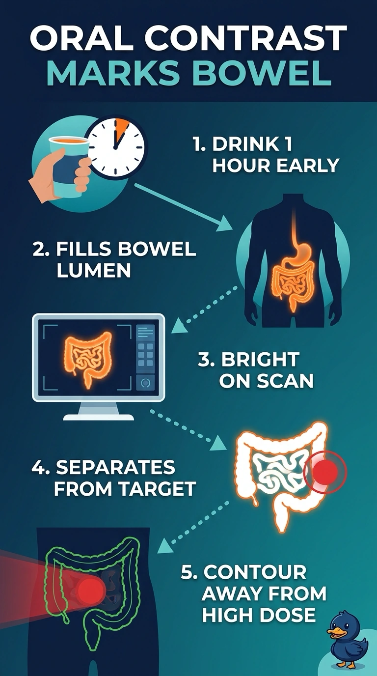
Oral contrast stays inside your gut, so it lights up the bowel lumen and lets you see where the loops sit beside the target. Intravenous contrast does a different job, enhancing structures such as tumors and lymph nodes that take up contrast from the blood. For your pelvic plan, bowel separation is the point of the drink.
Full Explanation
Positive oral contrast mixes with bowel contents and stays inside the lumen during the scan, producing a bright column that makes each loop easier to trace and contour as an organ at risk when it sits against the target. That separation is the purpose of the order and helps prevent bowel from being drawn into the high-dose region by mistake. Enhancing the tumor and lymph nodes is instead a role of intravenous contrast, which enters the bloodstream and is taken up by vascular tissue; an oral agent cannot sharpen nodal borders in the same way. Outlining the renal collecting system and ureters likewise depends on intravenous contrast that the kidneys excrete, serving urinary tract contouring rather than bowel separation. Defining the bladder wall is not the purpose of the oral dose; bladder assessment mainly relies on consistent filling and the natural soft-tissue contrast already present on CT. One exam caveat: oral contrast timing and concentration vary by protocol, so follow department policy rather than memorizing a schedule. Practical check: review the scan at the target level and confirm that bowel loops appear brighter than surrounding soft tissue before contouring.