
A patient with an akinetic inferior wall and a severely reduced ejection fraction is being evaluated for revascularization, and the cardiology team needs to know whether the resting defect is scar or hibernating myocardium. The nuclear medicine staff must pick a protocol that answers that viability question during a single visit. Which single-visit imaging protocol best answers the viability question?
Select an answer to reveal the explanation.
Short Explanation
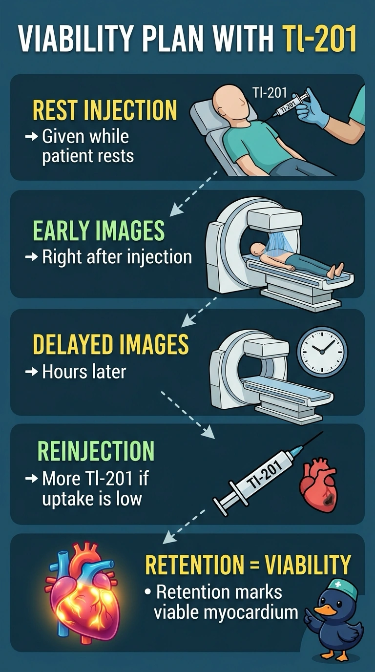
You're separating two look-alike problems, and that distinction decides the scan. Tracer that redistributes into a resting defect signals viable tissue, so redistribution is what helps distinguish hibernating muscle from scar. A single perfusion picture is like a photograph from one moment; it cannot show what happens afterward.
Full Explanation
Hibernating myocardium may have reduced resting perfusion but retain viable cells that take up Tl-201. Redistribution and, when needed, reinjection images acquired after the resting study can help distinguish viable tissue from scar, which generally shows little uptake. Reading viability from sestamibi reversibility alone fails because reversibility reports inducible ischemia, and a hibernating segment can look fixed on a rest-stress pair. Stopping with initial Tl-201 images fails because early perfusion pictures show tracer delivery and do not provide the later redistribution information. Judging viability from gated wall thickening and motion alone is unreliable because poor mechanical function does not by itself distinguish viable from scarred tissue. Exam caveat: other accepted viability approaches exist and facility protocols differ, so focus on the concept of tracer redistribution and retention rather than one camera setting. Practical check: before scheduling, confirm the protocol includes delayed or reinjection imaging, not just the resting acquisition.