
A quality review of a Tc-99m SPECT study attributes reduced contrast to photons scattered inside an average-size patient. The camera records events that deposit only part of the 140 keV photon energy and change direction. Which photon interaction in soft tissue best explains these scattered events?
Select an answer to reveal the explanation.
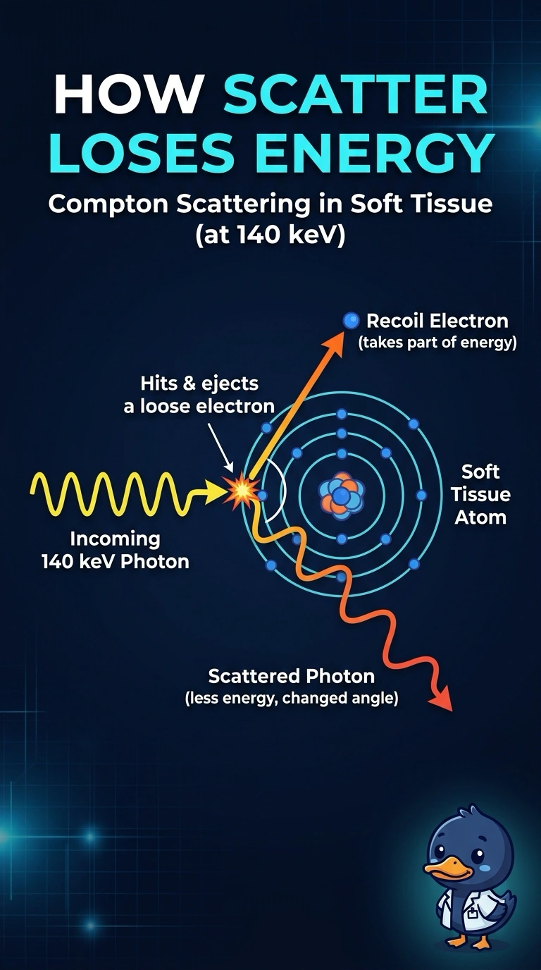
Short Explanation
Partial energy loss with a direction change is your clue: Compton scattering transfers some photon energy to a recoil electron while the photon continues at lower energy. A cue ball tapping another and rolling away slower shows the same split. Compton scattering is the interaction to name at 140 keV in tissue.
Full Explanation
Compton scattering in soft tissue best fits the observation: at 140 keV, an incident photon can transfer part of its energy to an electron and continue in a different direction at lower energy. The recoil electron deposits energy locally, while the scattered photon may escape the patient or reach the camera, contributing to detected scatter and reduced image contrast. Rayleigh (coherent) scattering is a real tissue-scatter process and changes photon direction, but it transfers negligible energy; it therefore does not explain events that deposit only part of the photon energy. In photoelectric absorption, the incident photon is absorbed rather than scattered. A fluorescent x-ray may escape after carrying away some energy, but that secondary emission does not make photoelectric absorption the interaction that scattered the original 140 keV photon. Compton scattering can also occur in the camera crystal and produce partial-energy events, but that location does not account for photons scattered inside the patient, as specified here. Exam caveat: partial-energy deposition alone is not unique to Compton scattering; identify where the interaction occurs and whether the outgoing photon is the scattered primary or a secondary emission. Practical check: review the energy-window and scatter-correction settings, and compare patient-scatter estimates with energy-window data before attributing a contrast loss solely to the interaction mechanism.