
A patient with a clear runny nose after a head injury only leaks fluid when bending forward, and the referring team wants the leak located before surgery. The department can give an intrathecal tracer and can return the patient for delayed imaging later the same day. Which imaging plan will best show where the cerebrospinal fluid is escaping?
Select an answer to reveal the explanation.
Short Explanation
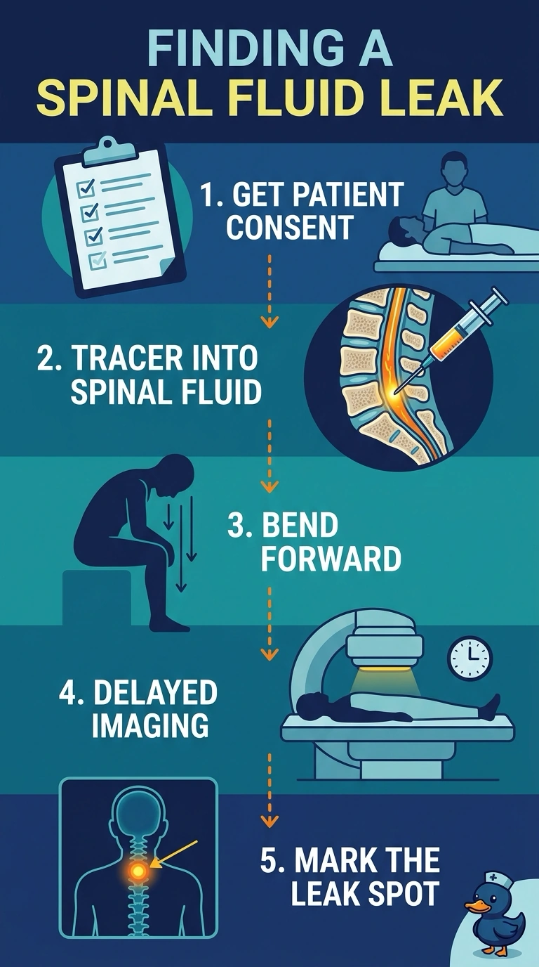
Because the leak shows up only when your patient leans forward, you need the tracer inside the cerebrospinal fluid and images that come back later, once it has had time to reach the nose. Going through a vein puts the tracer in the blood instead, and one early image set usually won't catch anything. It works like timing a delivery: right address, enough time for it to arrive.
Full Explanation
Cerebrospinal fluid moves slowly through the subarachnoid space, so a leak that only appears with bending needs two things at once: tracer placed directly in the spinal fluid and images taken later, once the tracer has had time to travel to the site of drainage. Bending forward is the provocative position that opens the leak, so activity collects where the fluid is escaping and shows up on delayed views. Giving the tracer by vein fails the route fact, because intravenously administered activity stays in the blood pool and never labels the cerebrospinal fluid. Imaging while the patient stays flat fails the positional fact, since the leak is intermittent and may not drain at all during those views. A single early image set fails the timing fact, because the tracer has not reached the nose when the first images are made. Exam caveat: exact imaging intervals and tracer choice follow the department's protocol and the prescriber's order, so learn your own lab's schedule rather than memorizing a universal one. Practical check: confirm consent, ask about prior reaction to the intrathecal agent, and place the patient in the posture that reproduces the leak before each delayed set.