
A four-year-old is booked for planar Tc-99m DMSA imaging to look for cortical scarring after a febrile urinary tract infection. The camera offers low-, medium-, and high-energy collimators, and the protocol calls for each kidney to be seen free of overlap from the spine, liver, and spleen. The child can lie still for one session only, so timing and views must be right the first time. Which acquisition plan fits this renal cortical study?
Select an answer to reveal the explanation.
Short Explanation
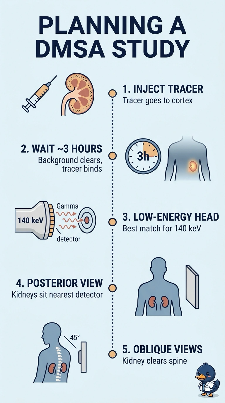
DMSA needs time to localize in the cortex while background activity clears, so images are acquired a few hours after injection. Match that delay with a low-energy collimator suited to technetium's 140 keV gamma, and add oblique views so each kidney clears the spine and the liver or spleen. It's like letting the water settle in a jar before you take the photo.
Full Explanation
Tc-99m DMSA localizes in the renal cortex, and activity in blood and soft tissue needs time to clear, which is why cortical images are collected about two to three hours after injection; an earlier view has higher background that can obscure a small scar. A low-energy collimator matches the 140 keV photopeak of technetium-99m and provides suitable septal geometry at that energy, while posterior and oblique projections help show each kidney clear of the spine and of the liver or spleen. Imaging immediately after injection is too early for this cortical study. A medium-energy collimator is intended for higher-energy photons and is not the appropriate choice for Tc-99m. Posterior and anterior views alone do not provide the oblique projections needed to separate the kidneys from overlapping structures. Uptake delays vary between protocols, so follow the department's written timing. Practical check: note the injection time, mount the low-energy collimator, and preview a static before committing to full counts.