
A SPECT study of the lumbar spine was acquired with the patient still, but the low count density and a sharp reconstruction filter leave the summed images grainy with speckled hot spots over the vertebral bodies. You need images reliable enough to judge focal uptake. Which reconstruction change should you make before reading this study?
Select an answer to reveal the explanation.
Short Explanation
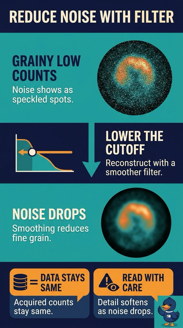
Lower the filter cutoff to use a smoother filter, which suppresses noise in these low-count images. It’s like softening a grainy photo: you may lose a little sharpness, but the vertebral uptake will be easier to judge.
Full Explanation
Lowering the reconstruction filter cutoff applies stronger smoothing, suppressing high-frequency noise that appears as grain and speckled hot spots in these low-count images. This makes focal uptake easier to assess, although it trades some spatial resolution for reduced noise. Increasing iterations and subsets can improve convergence and apparent detail, but in a low-count study it also tends to amplify statistical noise and can make the speckling more prominent; it does not address the stated image-quality problem. Attenuation correction with a registered transmission map is a standard way to compensate for photon loss and improve quantitative accuracy, but it does not remove count noise and may increase noise in corrected regions. Resolution-recovery modeling can improve apparent spatial resolution, yet it cannot substitute for noise suppression and may accentuate noisy fluctuations or produce reconstruction artifacts. Exam caveat: filter cutoff and the balance between noise and resolution depend on the acquisition, reconstruction method, and clinical task, so stronger smoothing is not universally preferable. Practical check: review the filtered images alongside the projection data and confirm that suspected focal uptake remains visible without excessive blurring.