
A four-year-old with known reflux is scheduled for cortical imaging to look for scarring. The images must separate small cortical defects from overlying ribs and keep bladder activity from clouding the lower poles. Which acquisition plan best meets those requirements?
Select an answer to reveal the explanation.
Short Explanation
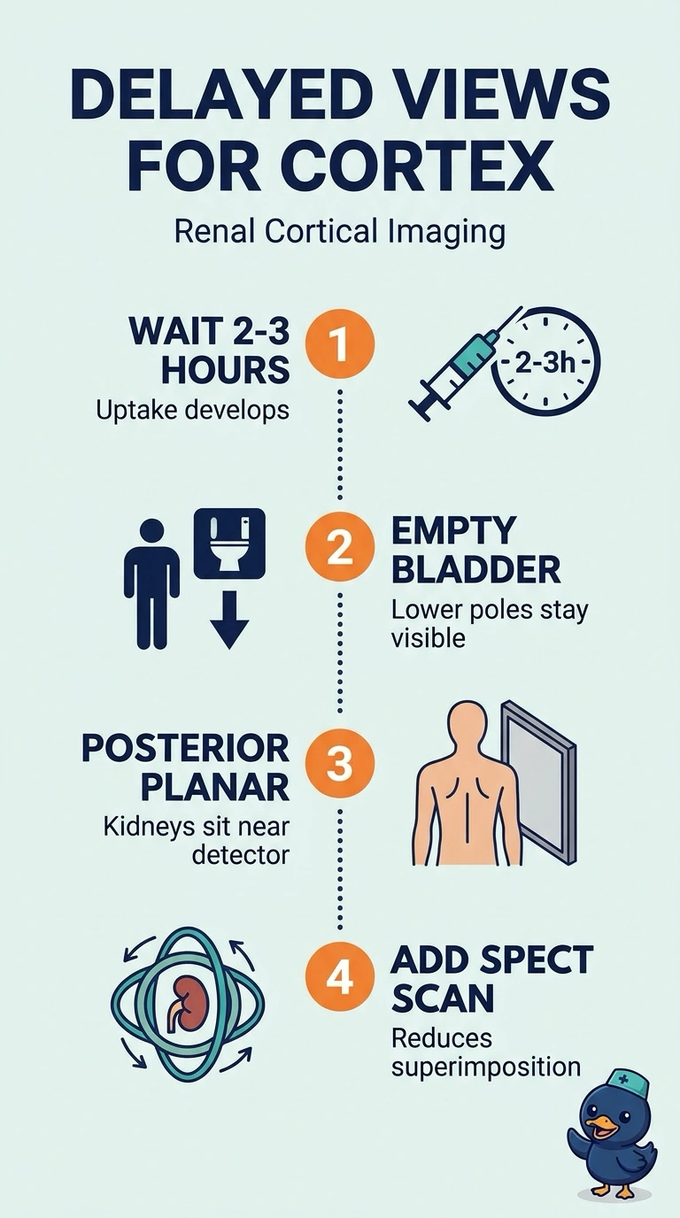
You’ll get the clearest view by waiting two to three hours after injection, then having the child void before posterior planar imaging and SPECT. Delayed imaging helps small cortical scars stand out, while SPECT separates them from overlapping ribs and an empty bladder keeps the lower poles clear.
Full Explanation
Tc-99m DMSA accumulates in proximal tubular cells, and the cortex-to-background ratio generally improves over the hours after injection. Delayed imaging allows cortical uptake to develop as circulating activity decreases, helping small cortical scars stand out. Posterior imaging places the kidneys closest to the collimator for good resolution, while SPECT provides tomographic localization that reduces superimposition from structures such as ribs. Having the child void before imaging removes a potential source of activity near the lower poles. Anterior and posterior planar images with SPECT obtained soon after injection include the needed views and bladder preparation, but imaging too early does not allow the usual delayed uptake to develop. Posterior planar images alone, even when delayed and obtained after voiding, do not provide tomographic separation of overlapping structures. Posterior planar images plus SPECT obtained without voiding provide the delayed and tomographic views, but bladder activity may cloud the lower poles. Caveat: the exact delay can vary by institutional protocol. Practical check: confirm that the child has voided and time the injection against the planned start of imaging.