
A child with a history of febrile urinary tract infection is scheduled for a Tc-99m dimercaptosuccinic acid study to look for a small cortical scar. The dose is drawn and the camera frees up right away, but images need low blood-pool activity, kidneys close to the detector, and an emptied bladder. Which acquisition plan best supports detection of that cortical defect?
Select an answer to reveal the explanation.
Short Explanation
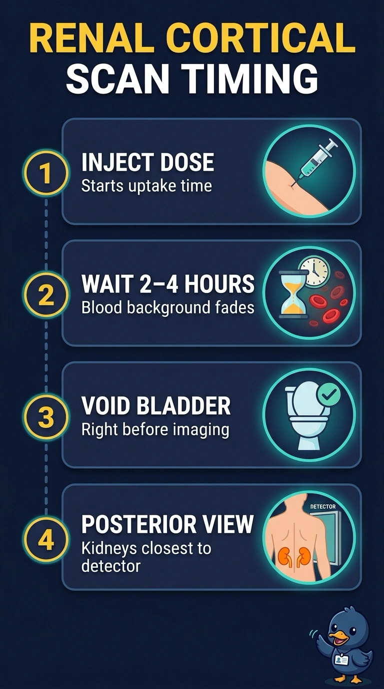
For the clearest view of a small cortical scar, you need delayed images—about two to four hours after voiding—with the kidneys close to the detector. It’s like letting background noise fade so a quiet signal stands out: delayed imaging improves contrast, while an early scan or a more distant kidney position can obscure the defect.
Full Explanation
Tc-99m DMSA localizes in the renal cortex, and delayed imaging allows background activity to clear, improving contrast for cortical defects. A small scar appears as a photopenic area against the normally active cortex. Keeping the kidneys close to the detector helps preserve spatial resolution, and voiding before imaging reduces activity in the bladder. Beginning a posterior static acquisition early leaves more background activity, while centering the kidneys in the field of view addresses coverage rather than contrast. Immediate SPECT also begins before background activity has cleared. Anterior imaging with a supine patient meets the delay and voiding requirements, but places the kidneys farther from the detector than posterior imaging does in that position. Imaging times can vary with the protocol; follow the applicable radiopharmaceutical labeling and local procedure. Document the injection time and void, then acquire images with the kidneys fully in the field.