
A patient treated for breast cancer asks why her physician ordered a nuclear medicine bone scan instead of another CT scan. Her history notes possible spread to several bones, and the order calls for a survey of the whole skeleton's activity. Which explanation correctly compares the whole-skeleton activity shown by the bone scan with the anatomy shown by CT?
Select an answer to reveal the explanation.
Short Explanation
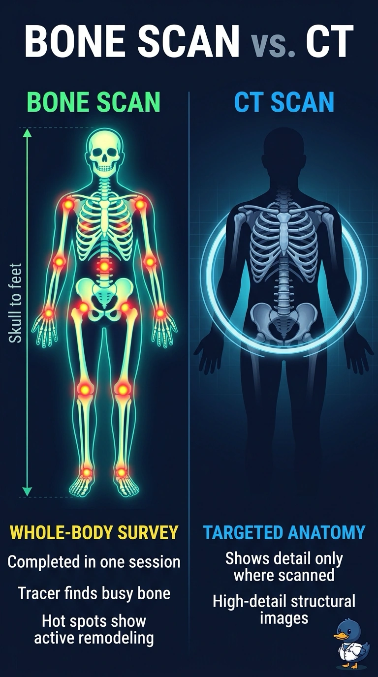
Here's the hook: the tracer accumulates where bone remodeling is increased, so a whole-body scan can flag abnormalities across the skeleton. A CT shows anatomy in the regions it images, so it answers anatomic rather than activity questions. That's the osteoblastic response working for you.
Full Explanation
The Tc-99m phosphonate tracer is cleared by the kidneys and adsorbs onto bone surfaces; uptake reflects local blood flow and osteoblastic activity. A whole-body planar acquisition can survey the skeleton from skull to feet in one session, which is useful when disease is suspected in several bones. A CT images the anatomic regions included in its scan and provides anatomic and density information rather than directly showing bone remodeling activity. A whole-body low-dose CT may cover much of the skeleton, but it does not provide the activity information requested here. Tracer uptake can be limited in some purely lytic lesions, so nuclear medicine findings are correlated with anatomic imaging. Confirm that the acquisition covers the intended skeletal regions.