
Indium-111 oxine-labeled autologous white blood cells are reinjected for a patient being evaluated for a suspected deep abdominal abscess, with imaging about a day later. Which mechanism carries the activity to the infected focus and causes it to accumulate there?
Select an answer to reveal the explanation.
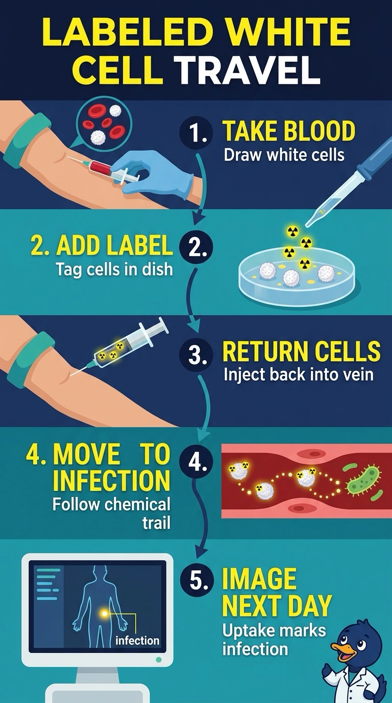
Short Explanation
After reinjection, your tagged white blood cells follow chemotaxis—like following a trail of signals—to the infection. They leave the bloodstream and gather at the infected focus.
Full Explanation
The correct mechanism is active recruitment of the reinjected, labeled autologous white blood cells: chemotactic signals draw them out of the circulation and into infected tissue, so the label accumulates with the cells. This is why indium-111 oxine leukocyte imaging can identify a deep abscess on delayed images. Gallium-67 citrate is also used to image infection, but its activity localizes through transport and binding involving transferrin and inflammatory proteins such as lactoferrin; it is not carried there by the patient’s reinjected, labeled leukocytes. FDG can show infection because activated inflammatory cells take up and phosphorylate the glucose analogue, trapping it intracellularly. That is metabolic uptake of a different tracer, not chemotactic trafficking of labeled white blood cells. Radiolabeled anti-granulocyte antibodies can target leukocytes at sites of infection, but they localize by binding cell-surface antigens rather than by labeling autologous cells that migrate in response to chemotaxis. Exam caveat: inflammation is not specific to infection, and normal or altered biodistribution can complicate interpretation. Practical check: confirm adequate cell labeling and review delayed images alongside the patient’s clinical findings and anatomic imaging.