
A 64-year-old with new exertional chest pain completes a two-day stress myocardial perfusion study. The raw data show sharp motion-free frames, and the lateral wall takes up less tracer at peak stress but fills in completely on the next morning's resting images. The cardiologist asks what that change means for the muscle. Which myocardial pathophysiology best explains the pattern?
Select an answer to reveal the explanation.
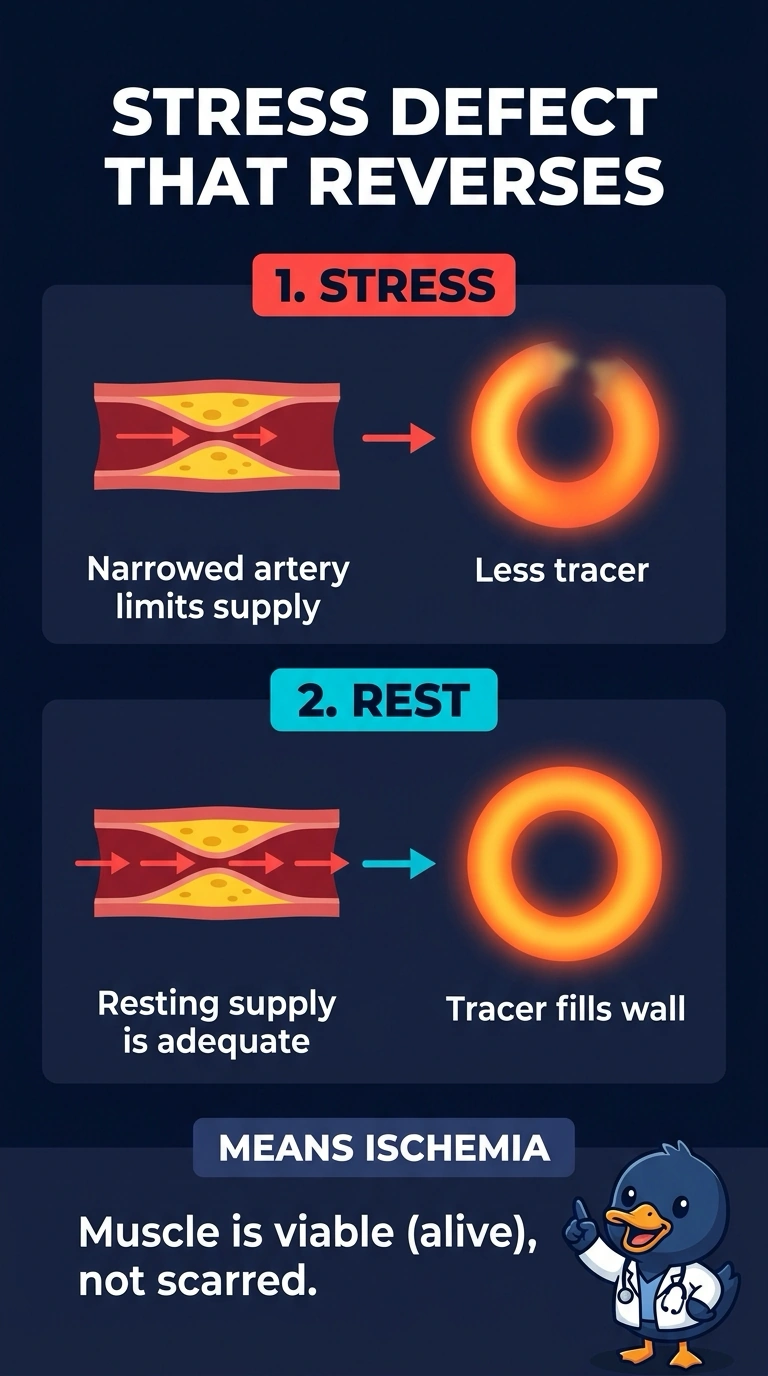
Short Explanation
When the lateral wall fills back in at rest, you’re seeing ischemia—living muscle that temporarily gets too little blood during exertion, like a road that backs up only at rush hour. Since it recovers at rest, it isn’t a fixed scar, and the sharp images make motion artifact less likely.
Full Explanation
A reversible perfusion defect is the classic scintigraphic pattern of stress-induced ischemia: at peak stress, blood flow may be insufficient to meet myocardial demand, so viable myocardium shows relatively reduced tracer uptake; at rest, perfusion may be adequate and uptake can appear normal. This pattern indicates viable tissue rather than a fixed scar. An old lateral-wall scar would generally remain a defect at rest, rather than filling in completely. Fixed soft-tissue attenuation with unchanged positioning would generally affect both acquisitions, not produce this reversible pattern. Stress-motion blur can cause apparently reduced uptake, but the stated clean, motion-free raw data make it less likely. An exam caveat is that attenuation correction or prone imaging can change a defect’s appearance, and differences in positioning between acquisitions can affect attenuation. As a practical check, review the projection movies and compare stress and rest images, including whether the defect follows a coronary territory.