
A patient with persistent staphylococcal bacteremia and new mid-back pain is being worked up for vertebral osteomyelitis. Imaging must show labeled white blood cell accumulation rather than bone turnover alone, normal vertebral marrow also avidly takes up labeled cells, and the suspected focus is small and deep in the spine. Which infection imaging plan best confirms the focus?
Select an answer to reveal the explanation.
Short Explanation
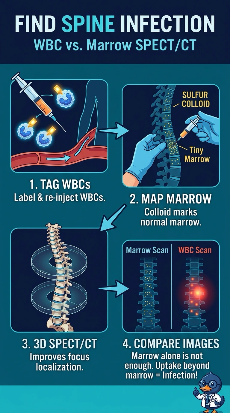
You picked the study that addresses both the marrow problem and the depth problem: compare labeled white blood cell uptake with the marrow distribution, using SPECT/CT to localize the focus.
Full Explanation
Vertebral osteomyelitis is a hard target because normal marrow and infection can both show uptake on a labeled white blood cell scan. A technetium-99m sulfur colloid scan maps functioning marrow; focal white blood cell uptake that does not correspond to colloid uptake supports infection rather than normal marrow. SPECT/CT helps localize a small deep focus in three dimensions. Gallium-67 citrate with colloid can be used in some marrow-infection evaluations, but it does not meet this scenario's requirement to demonstrate labeled white blood cell accumulation. Labeled white blood cell SPECT without a marrow comparison can leave normal vertebral marrow uptake ambiguous. Planar images with a colloid comparison do not provide the same three-dimensional localization of a small deep focus. Isotope choice and timing vary by protocol.