
A 66-year-old smoker with a left upper lobe mass and badly reduced lung capacity is being evaluated for surgery. The surgeon asks nuclear medicine for a number showing how much of the patient's total blood flow each lung contributes, so the resection can be planned safely. Which lung study plan best supplies that split-function result?
Select an answer to reveal the explanation.
Short Explanation
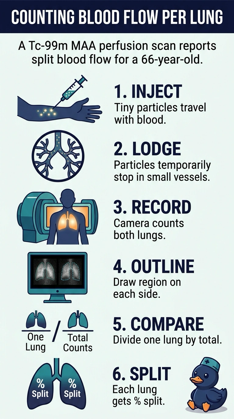
You went with perfusion, because split function is a blood-flow question and quantitative perfusion scintigraphy is what counts the tracer in each lung. Aerosol ventilation maps airflow instead, a visual read hands back a pattern rather than a number, and a first-pass study reports heart function. It's like judging two pipes by what arrives at each end instead of listening to the air move.
Full Explanation
Quantitative perfusion scintigraphy supplies the number the surgeon wants because technetium-99m macroaggregated albumin particles are too large to cross the pulmonary capillary bed, so each particle temporarily lodges downstream of the arteriole feeding that region in proportion to blood flow. After acquisition you place a region over each lung, subtract background, and compare counts, which yields a percentage of total flow for each side and lets the surgeon predict what remains after resection. An aerosol ventilation scan does produce regional information, but it describes airflow and gas distribution rather than perfusion, so it fails the blood-flow requirement. Reading the perfusion study visually for filling defects gives a pattern and a probability category instead of a numerical split, so it fails the quantitative requirement. A first-pass radionuclide angiocardiogram yields genuine numbers too, yet they describe cardiac output and ejection fraction, which is the wrong physiologic target. Exam caveat: region placement, decay correction, and camera geometry vary by protocol, so understand the principle rather than memorizing a counting recipe. Practical check: confirm the ordering indication, position the patient so both lung fields are complete on the image, and inspect the regions before reporting the percentages.