
A patient is 10 days after a total knee replacement with fever, a warm and swollen knee, and raised inflammatory markers. The surgeon wants to distinguish periprosthetic infection from mechanical loosening before revision work. Which nuclear medicine imaging plan is most appropriate?
Select an answer to reveal the explanation.
Short Explanation
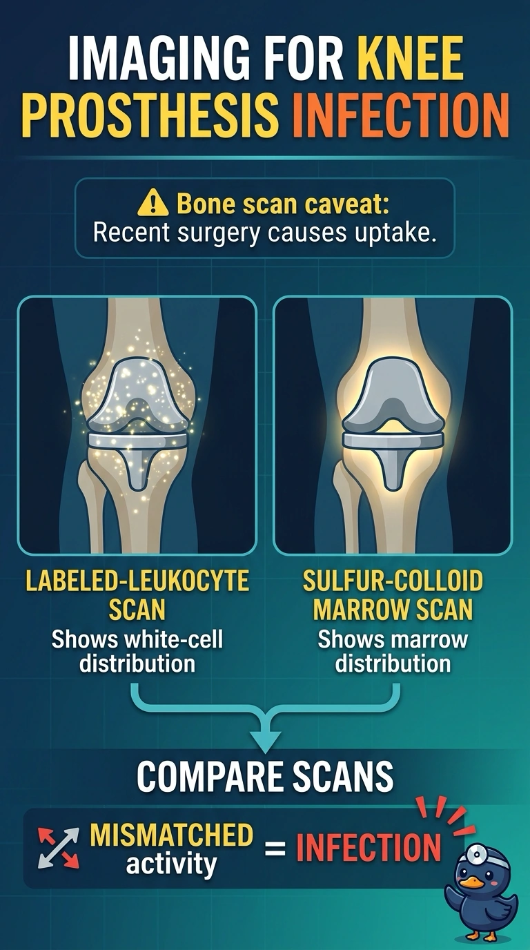
With fever and a warm, swollen knee so soon after surgery, you need imaging that helps separate infection from normal marrow activity around the prosthesis. Pair labeled-leukocyte imaging with sulfur-colloid marrow imaging for that comparison.
Full Explanation
Labeled-leukocyte imaging paired with sulfur-colloid marrow imaging is the most appropriate plan because it evaluates suspected infection while comparing leukocyte accumulation with the distribution of marrow activity. That comparison is particularly useful around a prosthesis, where marrow distribution may be altered and leukocyte uptake alone can be difficult to interpret. Labeled-leukocyte scintigraphy alone can localize a suspicious focus, but it lacks the complementary marrow study needed to help distinguish infection from normal or displaced marrow activity. Knee radiographs and metal-artifact-reduction CT can assess component position, fixation, and periprosthetic lucency, but they do not establish whether the febrile, inflamed knee is infected, so they do not answer the infection-versus-mechanical question. FDG PET/CT can show metabolically active inflammation, but only 10 days after surgery, normal healing and postoperative inflammation may also cause uptake, limiting its specificity for infection. An exam caveat is that inflammatory imaging findings can be nonspecific soon after arthroplasty, so postoperative timing matters when choosing and interpreting a study. In practice, confirm the exact postoperative interval and review the operative history and prior imaging with nuclear medicine before interpreting findings around the prosthesis.