
An adult with a dilated renal pelvis on ultrasound is scheduled for a diuretic renogram to separate true obstruction from a system that simply drains slowly. The patient arrives after a dry morning with a full bladder, and the order specifies a diuretic after peak uptake. Which preparation sequence will give the most interpretable washout curve?
Select an answer to reveal the explanation.
Short Explanation
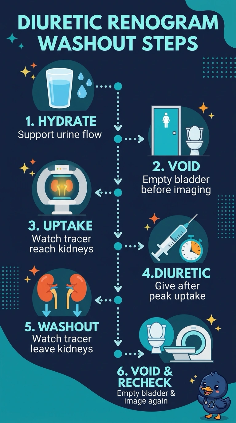
Hydrate steadily and have your patient void before imaging so a full bladder doesn’t slow drainage like a kink in a hose. Give the diuretic after peak uptake as ordered, then repeat imaging after they void to assess washout and residual activity.
Full Explanation
Hydrate the patient to support steady urine production, then have them void before imaging so a distended bladder is less likely to impede upper-tract drainage. After peak tracer uptake, administer the ordered diuretic to increase urine flow and challenge the collecting system; the resulting washout curve is more useful for distinguishing true obstruction from slow drainage. Repeat imaging after voiding: reduced residual activity may reflect improved drainage after bladder emptying, while persistent retention supports impaired washout. Giving the diuretic immediately after injection uses an early-diuretic approach rather than the specified post-peak timing; it changes the relationship between tracer uptake and the drainage challenge, making the requested protocol’s curve less interpretable. Imaging with voiding but no diuretic is a gravity/voiding-only assessment and omits the ordered pharmacologic challenge, so it cannot provide the intended diuretic washout assessment. Giving the diuretic without hydration may leave urine flow inadequate; slow clearance could then reflect low flow rather than obstruction. Exam caveat: diuretic timing varies among established renogram protocols, so no single timing should be treated as universal; follow the protocol specified for the study. Practical check: confirm adequate hydration, pre-imaging voiding, the ordered post-peak diuretic timing, and post-void imaging are all documented.