
A diuretic renogram is ordered for a mildly dilated collecting system found on a prior ultrasound. Post-furosemide images show slow clearance of tracer, yet from the first minutes the affected kidney showed weak cortical uptake and a flat, poorly rising curve. Which interpretation of the delayed drainage is best supported by this study?
Select an answer to reveal the explanation.
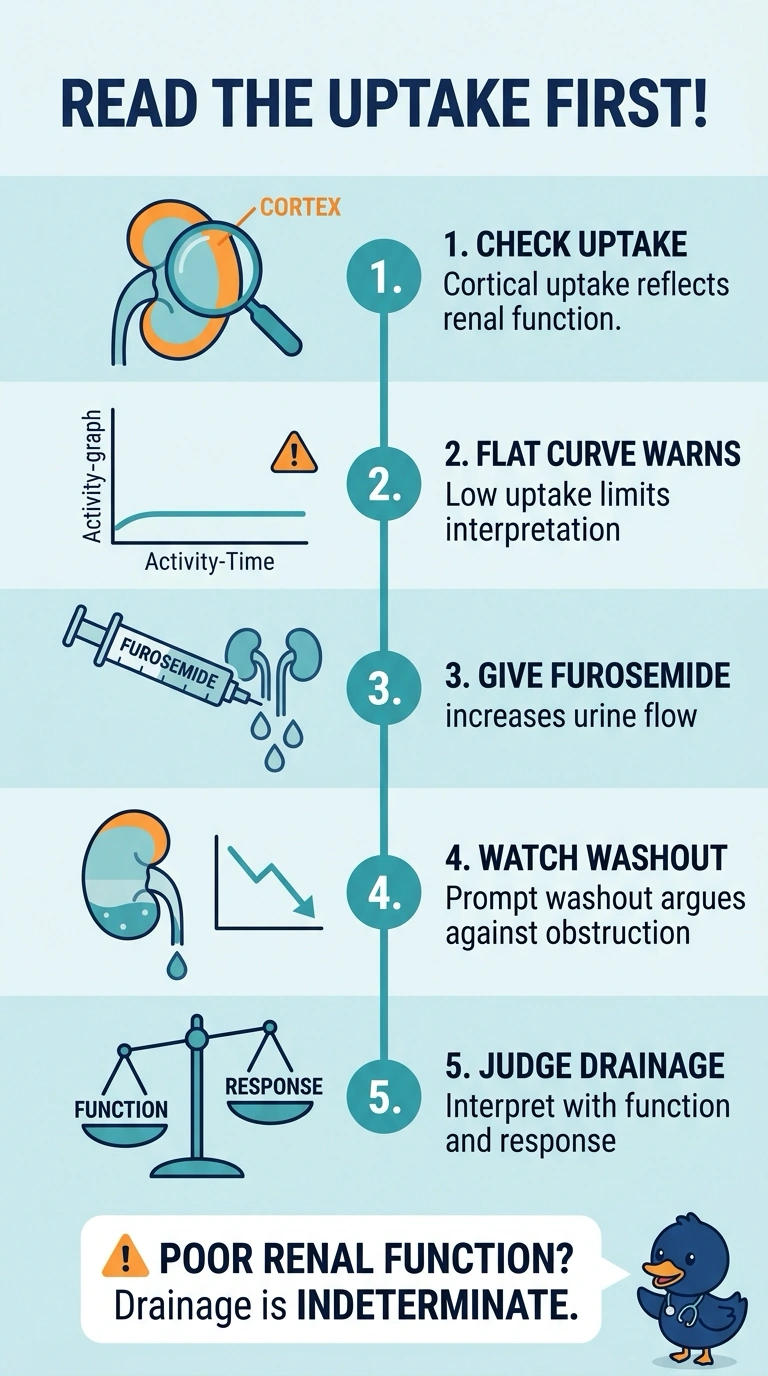
Short Explanation
Your drainage result is indeterminate because weak kidney function and poor tracer uptake can limit the response to furosemide. It’s like judging how quickly a bucket drains when the spigot barely opened—you can’t tell whether the drain is blocked.
Full Explanation
The best interpretation is indeterminate drainage. A diuretic renogram assesses whether tracer clears after furosemide, but that response depends on adequate delivery of tracer to the collecting system and sufficient renal function. Here, weak cortical uptake and a flat, poorly rising early curve indicate reduced function or poor tracer extraction, so slow apparent clearance cannot reliably distinguish obstruction from impaired renal handling. A prolonged post-furosemide half-time is commonly used to support obstruction, but treating it as decisive overlooks that poor function can prolong washout even without a mechanically significant blockage. Mild dilation can also behave as a compliant reservoir, and gravity-assisted or postvoid clearance can help identify nonobstructive stasis; however, that possibility does not justify calling this study nonobstructive without the relevant clearance findings, especially given the weak uptake. Persistent activity on supine and gravity-assisted images may strengthen concern for obstruction when renal function and tracer delivery are adequate, but those conditions are not established here, so that pattern cannot resolve this scan’s limitation. Exam caveat: a delayed drainage curve alone is not synonymous with obstruction; interpret it alongside uptake, curve shape, renal function, and postvoid images. Practical check: review differential function, image quality, hydration and bladder status, and gravity-assisted/postvoid drainage before recommending further anatomic evaluation or follow-up.