
A patient with new hydronephrosis has a dilated collecting system and mildly reduced kidney function, and the referral asks whether outflow is truly obstructed before surgery is considered. The team also wants a split-function number and a current clearance value. The patient can hydrate, void on command, and lie still for a dynamic run. Which renal study best separates obstruction from a system that drains slowly on its own?
Select an answer to reveal the explanation.
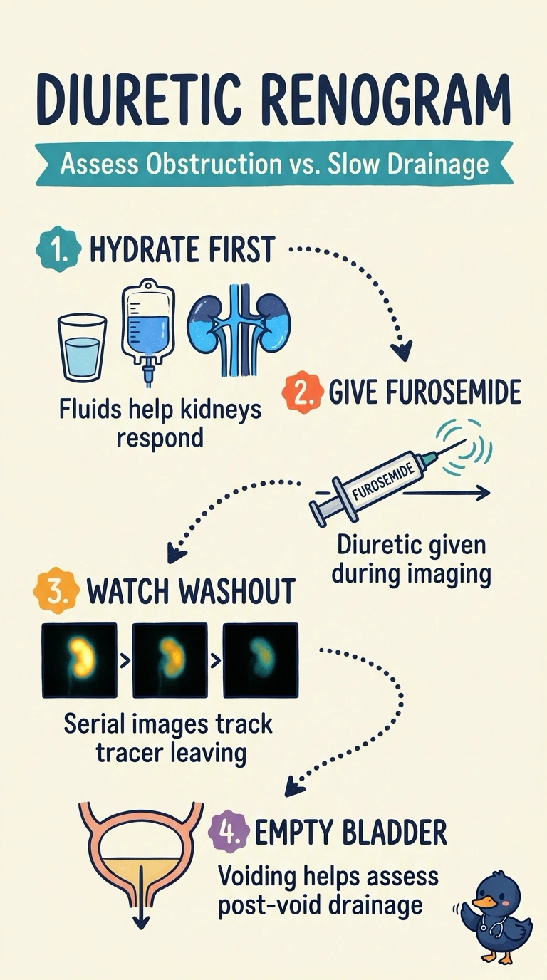
Short Explanation
To tell whether the collecting system is obstructed or simply drains slowly, you need to see how it responds when drainage is stimulated, so you choose a diuretic renogram. Furosemide turns the dynamic study into a washout curve, while a scan without the diuretic cannot provide the same drainage challenge. You also get differential function and tracer-clearance information.
Full Explanation
A diuretic renogram with dynamic imaging best addresses the question because it follows tracer through each kidney and the collecting system, then uses furosemide to challenge drainage. Persistent tracer retention despite diuresis supports functionally significant obstruction, whereas improved washout favors a dilated system that drains slowly without fixed obstruction. The study also provides differential renal function and quantitative tracer-clearance information. A MAG3 renogram without furosemide can show relative function and baseline transit, but without the diuretic challenge, slow emptying in a capacious collecting system may be difficult to distinguish from true obstruction. Functional MR urography can depict anatomy, drainage, and differential function, but does not provide the requested measured clearance value. DMSA cortical scintigraphy paired with measured GFR testing supplies cortical split function and clearance, but DMSA is not a dynamic drainage study and cannot establish whether the collecting system is obstructed. Exam caveat: delayed washout alone does not prove obstruction; hydration, bladder fullness, renal function, and region-of-interest selection affect interpretation. Practical check: confirm adequate hydration and have the patient void before imaging, then review the drainage curve together with postvoid images and differential function.