
A patient with a ventriculoperitoneal shunt has repeated headache and vomiting, and neurosurgery wants a study that shows cerebrospinal fluid still moving through the tubing into the abdominal cavity. The reservoir under the scalp can be tapped safely, and the team wants documentation of how quickly tracer travels the full length of the shunt. Which administration and imaging plan best demonstrates shunt patency?
Select an answer to reveal the explanation.
Short Explanation
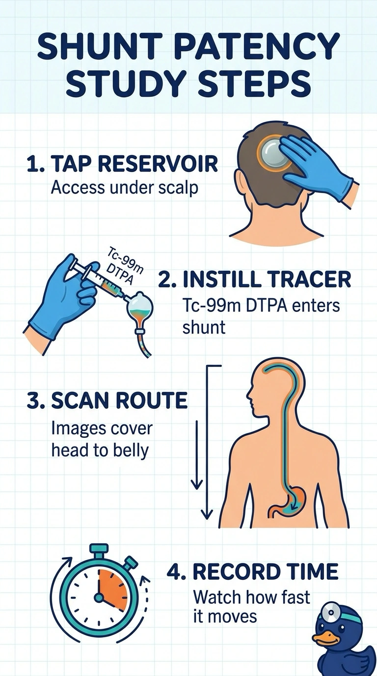
You'll tap the reservoir so the tracer starts inside the shunt, then follow it down to the belly with images taken in sequence. Serial views give you a shuntogram that shows both the route and the speed of flow.
Full Explanation
The correct plan introduces Tc-99m DTPA directly into the shunt reservoir, then follows its progression with serial imaging from the head through the distal tubing and into the peritoneal cavity. This documents both transit over time and passage along the full shunt course, the findings needed to assess patency in this scenario. Lumbar intrathecal administration can be used for studies of CSF flow, but it does not test flow from the shunt reservoir through the shunt itself; tracer reaching the abdomen by another route would not establish shunt patency. A single delayed whole-course image can show where tracer has accumulated, but cannot document how quickly it traveled or reliably distinguish delayed transit from an earlier pattern. Serial imaging only through the distal catheter tip can show tracer moving along the tubing, but does not document its entry or dispersal into the abdominal cavity, as requested. Exam caveat: protocols and acquisition intervals vary by institution, and a patent study does not exclude intermittent obstruction or explain every cause of symptoms. Practical check: confirm the reservoir injection site and image coverage with the nuclear medicine team, and correlate the observed transit and peritoneal activity with the clinical findings.