
During a myocardial perfusion study, the stress images show reduced tracer uptake limited to the anterior wall, and the rest images taken the next day show that same wall taking up tracer normally. Which combination of coronary supply and flow physiology explains this stress-to-rest difference?
Select an answer to reveal the explanation.
Short Explanation
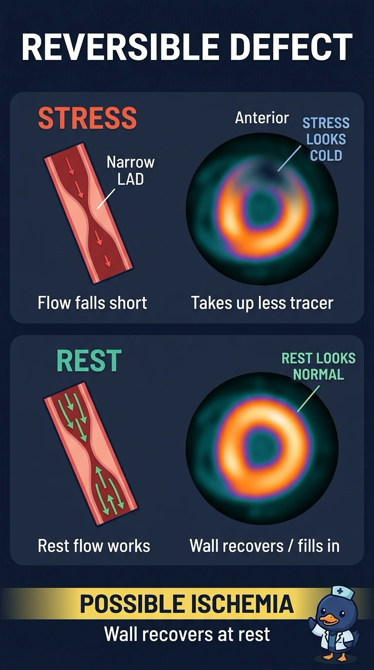
Your scan shows a reversible defect—like a road that handles light traffic but gets backed up at rush hour. The LAD supplies your anterior wall, and your resting flow is adequate even though stress flow can’t keep up with demand.
Full Explanation
A reversible defect is a typical imaging pattern of limited flow reserve: narrowing in the left anterior descending artery may allow adequate resting flow while preventing the increase in blood flow needed during stress. The anterior wall then receives less tracer relative to healthy segments and appears reduced, while uptake can appear normal at rest. An old infarct in the left anterior descending territory generally produces a fixed defect, with reduced uptake on both stress and rest images, although viable tissue around an infarct can show reversible ischemia. Breast attenuation can create an apparent anterior defect, but its effect is not necessarily identical between acquisitions because positioning and other imaging conditions can differ. Right coronary artery narrowing is not the expected supply explanation for reduced anterior-wall uptake; the anterior wall is usually supplied by the left anterior descending artery, which also supplies the anterior septum in most people. As an exam caveat, a reversible pattern supports ischemia but should be interpreted in light of possible attenuation and image quality. As a practical check, compare the stress and rest images side by side and confirm whether the defect resolves at rest.