
A 1.8 cm lesion is seen on ultrasound in the right lobe of the liver, and the radiologist has ordered a red blood cell blood-pool study to see whether it behaves like a hemangioma. The cells are labeled in vitro and reinjected, and the patient can come back later the same afternoon. Which timed imaging plan best characterizes this lesion?
Select an answer to reveal the explanation.
Short Explanation
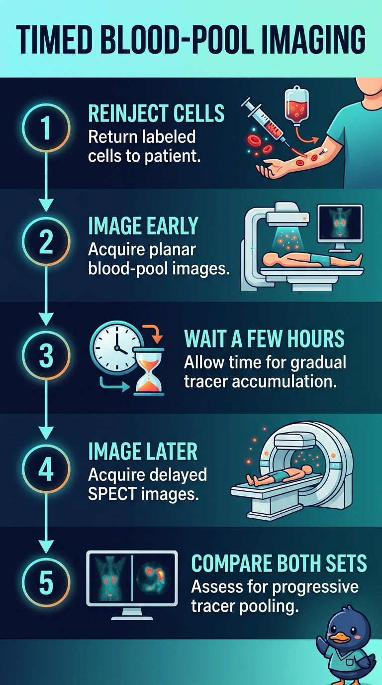
You’ll get early planar blood-pool images as a baseline, then delayed SPECT several hours later to catch a hemangioma slowly filling with labeled red cells—like watching a time-lapse. The delayed SPECT also helps pinpoint your small lesion within the liver.
Full Explanation
The correct plan pairs early planar blood-pool images with delayed SPECT several hours after reinjection. Early planar imaging documents the initial distribution of labeled red cells, while delayed tomographic imaging can show persistent blood-pool activity within a hemangioma and localize a small hepatic lesion more clearly than planar views alone. Early and delayed planar images are a reasonable way to assess changing activity, but they lack the spatial localization of SPECT and may not characterize a 1.8 cm lesion adequately. Early planar imaging followed by immediate SPECT includes the initial planar assessment, yet imaging before the delayed phase does not test for the characteristic persistent blood-pool pattern. Delayed planar imaging and SPECT provide the later tomographic assessment, but omitting early planar images removes the initial reference phase specified in the study plan. An exam caveat is that acquisition timing and protocols can vary by institution; the essential distinction here is early planar imaging versus delayed SPECT, not a universal number of hours. In practice, confirm the reinjection time and the department's RBC-labeling protocol, then schedule delayed SPECT within the requested same-afternoon window.